3 min
Study finds most cancer patients exposed to misinformation; UF researchers pilot 'information prescription'
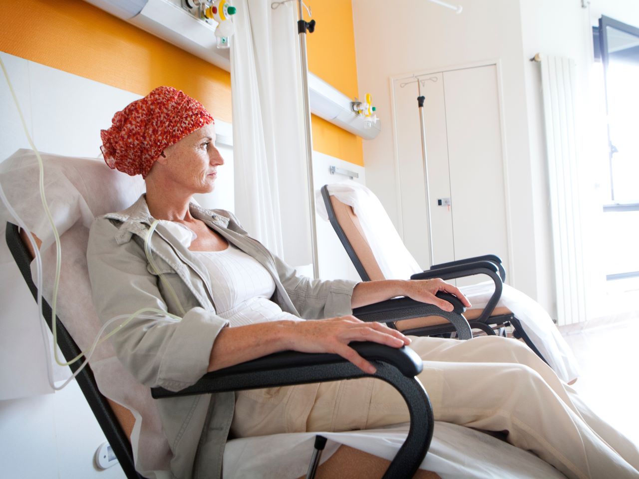
Ninety-three percent of patients with a new cancer diagnosis were exposed to at least one type of misinformation about cancer treatments, a UF Health Cancer Center study has found. Most patients encountered the misinformation — defined as unproven or disproven cancer treatments and myths or misconceptions — even when they weren’t looking for it. The findings have major implications for cancer treatment decision-making. Specifically, doctors should assume the patient has seen or heard misinformation. “Clinicians should assume when their patients are coming to them for a treatment discussion that they have been exposed to different types of information about cancer treatment, whether or not they went online and looked it up themselves,” said senior author Carma Bylund, Ph.D., a professor and associate chair of education in the UF Department of Health Outcomes and Biomedical Informatics. “One way or another, people are being exposed to a lot of misinformation.” Working with oncologists, Bylund and study first author Naomi Parker, Ph.D., an assistant scientist in the UF Department of Health Outcomes and Biomedical Informatics, are piloting an “information prescription” to steer patients to sources of evidence-based information like the American Cancer Society. The study paves the way for other similar strategies. Most notably, the study found the most common way patients were exposed to misinformation was second hand. “Your algorithms pick up on your diagnosis, your friends and family pick up on it, and then you’re on Facebook and you become exposed to this media,” Parker said. “You’re not necessarily seeking out if vitamin C may be a cure for cancer, but you start being fed that content.” And no, vitamin C does not cure cancer. Health misinformation can prevent people from getting treatment that has evidence behind it, negatively affect relationships between patients and physicians, and increase the risk of death, research has shown. People with cancer are particularly vulnerable to misinformation because of the anxiety and fear that comes with a serious diagnosis, not to mention the overwhelming amount of new information they have to suddenly absorb. While past research has studied misinformation by going directly to the source — for instance, studying what percentage of content on a platform like TikTok is nonsense — little research has looked at its prevalence or how it affects people. The team first developed a way to identify the percentage of cancer patients exposed to misinformation. UF researchers collaborated with Skyler Johnson, M.D., at Huntsman Cancer Institute, an internationally known researcher in the field. The survey questions were based on five categories of unproven or disproven cancer treatments — vitamins and minerals, herbs and supplements, special diets, mind-body interventions and miscellaneous treatments — and treatment misconceptions. The myths and misconceptions were adapted from National Cancer Institute materials and included statements like “Will eating sugar make my cancer worse?” The team surveyed 110 UF Health patients diagnosed with prostate, breast, colorectal or lung cancer within the past six months, a time when patients typically make initial treatment decisions. Most had heard of a potential cancer treatment beyond the standard of care, and most reported they had heard of at least one myth or misconception. The most common sources were close friends or family and websites, distant friends/associates or relatives, social media and news media. The findings mark a shift in misinformation research, with major implications for the doctor-patient relationship, said Bylund, a member of the Cancer Control and Population Sciences research program at the UF Health Cancer Center. “I still think media and the internet are the source and why misinformation can spread so rapidly, but it might come to a cancer patient interpersonally, from family or friends,” she said. Most patients rarely discussed the potential cancer treatments they had heard about with an oncologist, the study also found. Next, the researchers plan to survey a wider pool of patients, then study the outcomes of interventions designed to decrease misinformation exposure, like the information prescription.