Experts Matter. Find Yours.
Connect for media, speaking, professional opportunities & more.
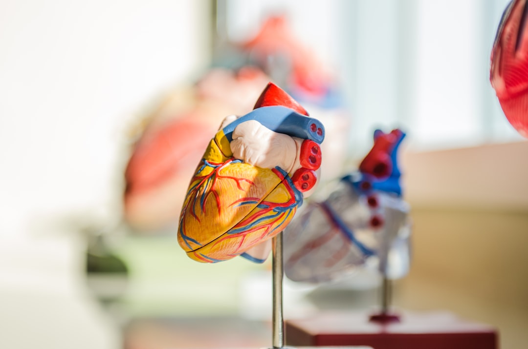
Heart valve developed at UC Irvine shines in early-stage preclinical testing
UC Irvine researchers designed and developed a minimally invasive replacement pulmonary heart valve. Created for pediatric patients, the device can be expanded as children grow, eliminating the need for multiple surgeries. The team successfully conducted laboratory and early-stage animal feasibility testing of the implant, crucial steps toward approval for human use. Irvine, Calif., June 23, 2025 — Researchers at the University of California, Irvine have successfully performed preclinical laboratory testing of a replacement heart valve intended for toddlers and young children with congenital cardiac defects, a key step toward obtaining approval for human use. The results of their study were published recently in the Journal of the American Heart Association. The management of patients with congenital heart disease who require surgical pulmonary valve replacement typically occurs between the ages of 2 and 10. To be eligible for a minimally invasive transcatheter pulmonary valve procedure, patients currently must weigh at least 45 pounds. For children to receive minimally invasive treatment, they must be large enough so that their veins can accommodate the size of a crimped replacement valve. The Iris Valve designed and developed by the UC Irvine team can be implanted in children weighing as little as 17 to 22 pounds and gradually expanded to an adult diameter as they grow. Research and development of the Iris Valve has been supported by the Eunice Kennedy Shriver National Institute of Child Health and Human Development; the National Heart, Lung, and Blood Institute; and the National Science Foundation. This funding has enabled benchtop fracture testing, which demonstrated the valve’s ability to be crimped down to a 3-millimeter diameter for transcatheter delivery and subsequently enlarged to 20 millimeters without damage, as well as six-month animal studies that confirmed successful device integration within the pulmonary valve annulus, showing valve integrity and a favorable tissue response. “We are pleased to see the Iris Valve performing as we expected in laboratory bench tests and as implants in Yucatan mini pigs, a crucial measure of the device’s feasibility,” said lead author Arash Kheradvar, UC Irvine professor of biomedical engineering. “This work represents the result of longstanding collaboration between our team at UC Irvine and Dr. Michael Recto at Children’s Hospital of Orange County built over several years of joint research and development.” Congenital heart defects affect about 1 percent of children born in the United States and Europe, with over 1 million cases in the U.S. alone. These conditions often necessitate surgical interventions early in life, with additional procedures required to address a leaky pulmonary valve and prevent right ventricular failure as children grow. The Iris Valve can be implanted via a minimally invasive catheter through the patient’s femoral vein. The Kheradvar group employed origami folding techniques to compress the device into a 12-French transcatheter system, reducing its diameter to no more than 3 millimeters. Over time, the valve can be balloon-expanded up to its full 20-millimeter diameter. This implantation method, along with the ability to begin treatment earlier in very young patients, helps mitigate the risk of complications from delayed care and reduces the need for multiple surgeries in this vulnerable population. “Once the Iris Valve comes to fruition, it will save hundreds of children at least one operation – if not two – throughout the course of their lives,” said Recto, an interventional pediatric cardiologist at CHOC who’s also a clinical professor of pediatrics at UC Irvine. “It will save them from having to undergo surgical pulmonary valve placement, as the Iris Valve is delivered via a small catheter in the vein and can be serially dilated to an adult diameter and also facilitate the future placement of larger transcatheter pulmonary valves – with sizes greater than 20 millimeters, like the Melody, Harmony and Sapien devices – if needed.” Kheradvar said that the next phase of preclinical testing of the Iris Valve is funded by the Brett Boyer Foundation, which is committed to supporting research into treatments for congenital heart disease. “We are actively engaged with the U.S. Food and Drug Administration to define and carry out the required experiments and documentation for first-in-human authorization of the Iris Valve,” Kheradvar said. “Our team is urgently advancing the Iris Valve through preclinical studies to enable its clearance for first-in-human use. This is a critical step toward providing toddlers – who currently have no viable minimally invasive treatment until they reach the 45-pound threshold – with a much-needed option.” First co-author Nnaoma Agwu, a biomedical engineering Ph.D. candidate at UC Irvine, said: “The development of the Iris Valve required a strong and knowledgeable team that understood the clinical and mechanical design requirements. This accomplishment would not have been possible without the collaboration of talented clinicians, veterinarians and engineers. With this milestone reached, we are rigorously advancing the Iris Valve’s development, setting our sights on human clinical trials.” Joining Kheradvar, Recto and Agwu as co-authors of the article in Journal of the American Heart Association were Daryl Chau, a recent UC Irvine master’s graduate; Gregory Kelley and Tanya Burney, both research specialists at UC Irvine, with Burney also affiliated with the Beckman Laser Institute; Ekaterina Perminov, a clinical veterinarian with UC Irvine’s University Laboratory Animal Resources; and Christopher Alcantara, a radiology technician at CHOC. About UC Irvine’s Brilliant Future campaign: Publicly launched on Oct. 4, 2019, the Brilliant Future campaign aims to raise awareness and support for the university. By engaging 75,000 alumni and garnering $2 billion in philanthropic investment, UC Irvine seeks to reach new heights of excellence in student success, health and wellness, research and more. The Samueli School of Engineering plays a vital role in the success of the campaign. Learn more by visiting https://brilliantfuture.UC Irvine.edu/the-henry-samueli-school-of-engineering About the University of California, Irvine: Founded in 1965, UC Irvine is a member of the prestigious Association of American Universities and is ranked among the nation’s top 10 public universities by U.S. News & World Report. The campus has produced five Nobel laureates and is known for its academic achievement, premier research, innovation and anteater mascot. Led by Chancellor Howard Gillman, UC Irvine has more than 36,000 students and offers 224 degree programs. It’s located in one of the world’s safest and most economically vibrant communities and is Orange County’s second-largest employer, contributing $7 billion annually to the local economy and $8 billion statewide. For more on UC Irvine, visit www.uci.edu. Media access: Radio programs/stations may, for a fee, use an on-campus studio with a Comrex IP audio codec to interview UC Irvine faculty and experts, subject to availability and university approval. For more UC Irvine news, visit news.uci.edu. Additional resources for journalists may be found at https://news.uci.edu/media-resources.


In the news: Chronicle features University of Delaware's Career Center
At the University of Delaware, career-development officials believe in teaching students how networking exists all around them, in both curricular and co-curricular realms, by taking career readiness outside the Career Center and infusing networking principles and practices into academic courses, social activities and alumni events. UD's Career Center was highlighted in a recent Chronicle of Higher Education article for innovative and exemplary networking practices and teachings. “If we get students to not think about networking as this static skill I have to build, and it’s more of a natural part of who I am, and it’s in my toolbox, it becomes less arduous, less scary, and easier to do," Rachel Coppola, UD’s Director of Life Design and Career Integration, said in the featured video. Reporters wishing to speak to career experts can reach out to mediarelations@udel.edu.


Why College Students Are Storming Fields More Often
In his most recent Forbes article, Dr. Marshall Shepherd takes a scientific look at why college students and fans storm football fields, blending insights from psychology, meteorology, and social dynamics. He explains that field-storming is not simply a burst of emotion—it’s a predictable outcome of collective excitement and shared identity. After an unexpected win or a high-stakes rivalry game, thousands of people simultaneously experience what psychologists call “emotional contagion,” amplifying feelings of unity and celebration. This shared surge, combined with environmental cues like stadium acoustics and crowd density, transforms the act into what Shepherd calls a form of “social weather event.” “Storming the field isn’t chaos—it’s choreography fueled by emotion and crowd physics.” Shepherd also examines the logistical and safety implications. He notes that while universities often celebrate these spontaneous displays of school pride, they carry risks ranging from crowd injuries to property damage. Yet, institutions are reluctant to ban them outright because these moments reinforce fan loyalty and media attention. Shepherd suggests that the solution lies in better understanding crowd behavior: designing stadiums with safe egress routes, training security teams to manage surges, and anticipating emotional tipping points rather than reacting afterward. “Understanding the science behind fan behavior lets us manage energy, not suppress it.” Ultimately, Shepherd’s piece reframes field-storming as a fascinating mix of culture and physics—where joy, identity, and momentum collide. He urges universities to see these moments not as mere rule-breaking but as opportunities to study human behavior in motion, and to design environments that celebrate passion without compromising safety. Dr. J. Marshall Shepherd is a leading international weather-climate expert and is the Georgia Athletic Association Distinguished Professor of Geography and Atmospheric Sciences at the University of Georgia. Dr. Shepherd was the 2013 President of American Meteorological Society (AMS), the nation’s largest and oldest professional/science society in the atmospheric and related sciences. View his profile here Dr. J. Marshall Shepherd is available to speak with the media about this interesting topic - simply click on his icon now to arrange an interview today.

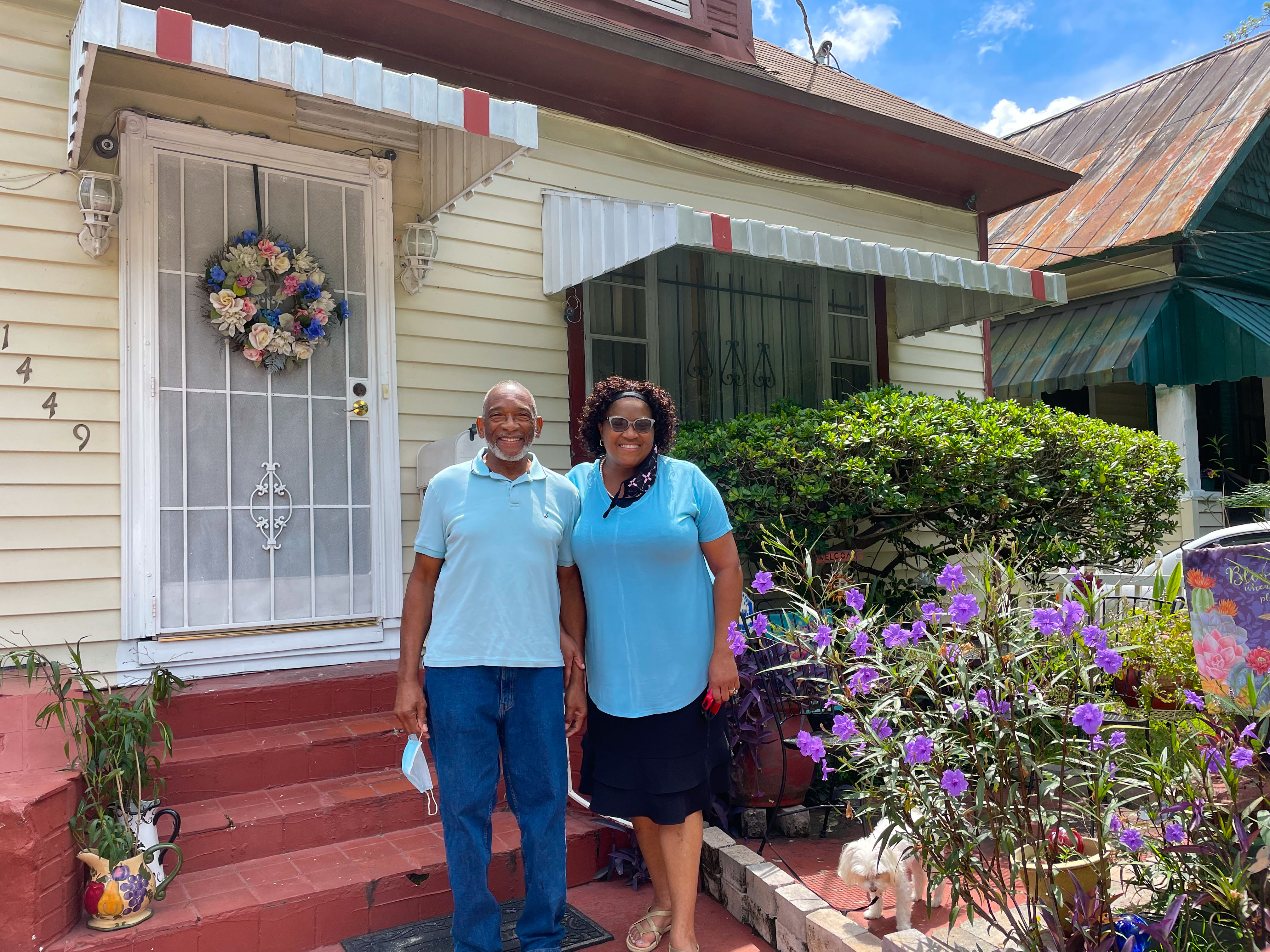
UF builds community resilience in Jacksonville’s Historic Eastside neighborhood
As the University of Florida continues to expand its presence in Jacksonville, Gators are undertaking sustainability projects to improve the city’s neighborhoods. Faculty and students in the College of Design, Construction and Planning’s Florida Institute for Built Environment Resilience (FIBER) have spent the past four years focusing on the role of housing design in community health resilience in Jacksonville’s Historic Eastside neighborhood, interviewing resident stakeholders and collaborating with citywide organizations that are helping to restore older homes. Findings from the UF research will be instrumental in informing future community planning and housing design decisions, potentially leading to more health-centered, sustainable neighborhoods. “Our research in Jacksonville focuses on how we can inform the development of community infrastructure that holistically supports human well-being across mental, emotional, and physical dimensions,” said Lisa Sundahl Platt, Ph.D., a FIBER research faculty member and an assistant professor of interior design at UF, who added that this holistic, health-centered approach is known as salutogenic design. “We are also actively collaborating with community organizations in Jacksonville and researchers from UF to explore improved strategies for designing and constructing community infrastructure that effectively responds to potential hazards.” A community-wide collaboration UF has conducted a pilot study over the past year on the Jacksonville-based Restore, Repair, and Resilience (R3) initiative that is underway in Historic Eastside – surveying residents about how the design quality of their housing and surrounding environments affects their overall well-being. This interdisciplinary project has brought together FIBER and members of the R3 Group – a coalition of organizations that includes the JEA utility company, LIFT JAX (committed to eradicating generational poverty), the Historic Eastside Community Development Corporation, the United Way of Northeast Florida, and Local Initiatives Support Corporation Jacksonville. FIBER-led research has received ongoing support from the Florida Resilient Cities grant, which is funded by the Jessie Ball duPont Fund. The scope of the R3 project is being scaled up through a U.S. Department of Energy grant awarded through JEA, which will allow for an expansion of home revitalization efforts in Eastside Jacksonville. FIBER’s ongoing housing and health community action research on these efforts will be supported through a grant from the LS3P Foundation. “Many residences we evaluated need help with improvements to housing energy efficiency, building ventilation, building shell structural integrity, and materiality,” Platt said. “For example, underperforming flooring material can create potential trip hazards for older adults. Deterioration in interior materials, caused by degrading components of the building envelope, can also lead to mold and mildew growth in interior environments, which can contribute to poor interior environmental quality issues and acute and chronic health conditions.” Respiratory health issues are often caused by material and ventilation design failures, which can affect people of all ages, especially vulnerable populations such as children and older adults. Oftentimes, interior designers see that the environmental risks that compromise human well-being are coming from both the outside and inside of the buildings. “As we continue to address priorities, our focus extends beyond energy and building efficiency to encompass comprehensive factors of built environment resilience that impact overall community health and well-being,” Platt said. “There's still significant progress to be made in the design of sustainable housing that supports community salutogenic health." Keeping residents safe and healthy UF research has continued to prove that interior resilience for living environments plays a vital role in people’s mental and physical health. “People spend roughly 90% of their time indoors, so it is important to understand the types of design conditions and materials that we’re putting into spaces and how they can affect the occupants of those living in said spaces,” said UF student Milena Rodriguez Mendez, who is one of Platt’s graduate research assistants. Students like Mendez are using qualitative and quantitative research methods to engage in collaborative community-led research that includes academics, for-profit organizations, nonprofits, citizen scientists, and neighborhood stakeholders. “I aim to center my work on social justice and equity, and I believe this initiative represents a meaningful step in that direction,” Mendez said. “Our focus is on the residents of this vibrant yet at-risk community.” FIBER researcher Jason von Meding added, “We want to know how future housing policies can address some future health concerns. We have a lot of youth in the community that are participating, which I think is important.” The FIBER housing and health team is actively pursuing additional funding to expand this research, in collaboration with UF Health Jacksonville’s Department of Community Engagement. “Our goal is to develop an open-source online platform that disseminates lessons learned and proof-of-concept findings on the impact of regenerative housing design on human and ecological health,” Platt said. “This resource will be valuable for other cities and neighborhoods facing similar challenges in housing quality, affordability, and accessibility.” Looking to know more about this project or connect with Lisa Platt? Simply click on her icon now to arrange an interview today.


Multi-university AI research may revolutionize wildfire evacuation
As wildfires grow wilder, the University of Florida and two other universities are developing large language models to make evacuations safer and more efficient. Armed with a nearly $1.2 million National Science Foundation grant, UF, Johns Hopkins University and the University of Utah are creating these AI-based models to simulate human behavior during evacuations – information that will help emergency managers shape more effective evacuation plans. “Strengthening wildfire resilience requires accurate modeling and a deep understanding of collective human behavior during evacuations,” said UF project lead Xilei Zhao, Ph.D., an associate professor with the Engineering School of Sustainable Infrastructure and Environment. “There is a critical need for simulation models that can realistically capture how civilians, incident commanders and public safety officials make protective decisions during wildfires.” Xilei Zhao focuses on developing and applying data and computational science methods to tackle problems in transportation and resilience. View her profile here Existing simulation models face limitations, particularly with reliable predictions under various wildfire scenarios. New AI models can simulate how diverse groups of people behave and interact during the hurried scramble to seek safety. Zhao’s team is developing a convergent AI framework for wildfire evacuation simulations powered by psychological theory-informed large language models. The project will produce simulation methods to promote teaching, training and learning, and support wildfire resilience by allowing public safety officials to use open-access tools. “This research seeks to be a transformative step toward improving the behavioral realism, prediction accuracy and decision-support capability of wildfire evacuation simulation models,” Zhao said. Zhao partnered with John Hopkins professor Susu Xu, Ph.D., and University of Utah professors Thomas Cova, Ph.D., and Frank Drews, Ph.D. The preliminary results of the study were recently presented at the 63rd Annual Meeting of the Association for Computational Linguistics. “In that paper, we started to train the model on the survey data we collected to see how we can accurately predict people's evacuation decisions with LLMs,” Zhao said. Research objectives include extending the Protective Action Decision Model for civilians and public safety officials, developing psychological theory-informed large language model agents for protective modeling and generating a realistic synthetic population as input for the simulation platform. The team also plans to develop learning-based simulations and predict human behavior under scenarios such as fire spread, warning and infrastructure damage. This research comes at a critical time, as the number of wildfires has significantly increased globally. About 43% of the 200 most damaging fires occurred in the last decade leading up to 2023, according to a recent study in Science. The intensity, size and volume of wildfires are threatening more urban areas. “If you go into the urban area, many people do not have cars, or they need additional mobility support,” Zhao said. “For example, the LA fires impacted nursing homes with a lot of elderly people, many of whom are immobile or lack the ability to drive. That's a big problem. This would be very relevant to them.” The large language models will provide important context for evacuation planning as well as real-time decision making. “We envision this tool being used during planning,” Zhao said, “so emergency managers can test different kinds of scenarios to determine how to draw the evacuation zones, where to issue the orders first and how to design the communications messaging.” This is important research and critical as wildfires become more common across North America. If you're a reporter looking to connect and learn more - then let us help. Xilei Zhao is available to speak with media - simply click on her icon now to arrange an interview today.


LSU Launches Louisiana’s Most Advanced Microscope at Research Core Facility
LSU’s Advanced Microscopy and Analytical Core (AMAC) facility gives Louisiana researchers access to 16 state-of-the-art instruments, including a new Spectra 300 Scanning Transmission Electron Microscope (S/TEM) for atomic-scale imaging and analysis. The new microscope—the most advanced in Louisiana—was installed with $10 million in support from the U.S. Army. Standing almost 13 feet tall on a platform isolated from vibration, the S/TEM required major renovations, including a raised ceiling, acoustic wall panels, and a magnetic field cancellation system to ensure the instrument’s stability and performance. The microscope offers magnification up to 10 million times, powerful enough to enlarge a single grain of Mississippi River silt to the size of Tiger Stadium. “This is a transformational moment for LSU and for the future of research in Louisiana,” Interim LSU President Matt Lee said. “With the installation of the most advanced microscope in the state, LSU is once again demonstrating how we’re delivering on our promises—leading in research, innovation, and service to the state and nation.” The launch of the AMAC and S/TEM demonstrates LSU’s increased investment in providing its faculty and partners with the best possible equipment for research and discovery, including for national defense, energy, and health. “Winning in research is no different than winning in athletics—the best facilities attract the best talent, and you need the best of both to win,” LSU Vice President of Research and Economic Development Robert Twilley said. “Today’s launch is about a state-of-the-art microscope but also the launch of the AMAC as our first research core facility at LSU—the first of more to come to attract, train, and supply the best research talent for Louisiana and build research teams that win.” Using a finely focused electron beam and techniques such as energy dispersive X-ray spectroscopy (EDS) and electron energy loss spectroscopy (EELS), the S/TEM can reveal both structure and chemistry at atomic resolution. These capabilities drive advances in materials science—improving semiconductors, solar cells, batteries, catalysts, coatings, and alloys—while supporting biomedical research by mapping drug delivery, uncovering the structures of viruses and bacteria, and improving medical implant design. LSU’s AMAC research core facility was recently rebranded, changing its name from the Shared Instruments Facility (SIF). Learn more about how AMAC instruments help unlock millions in federal research funding to Louisiana and deliver solutions.


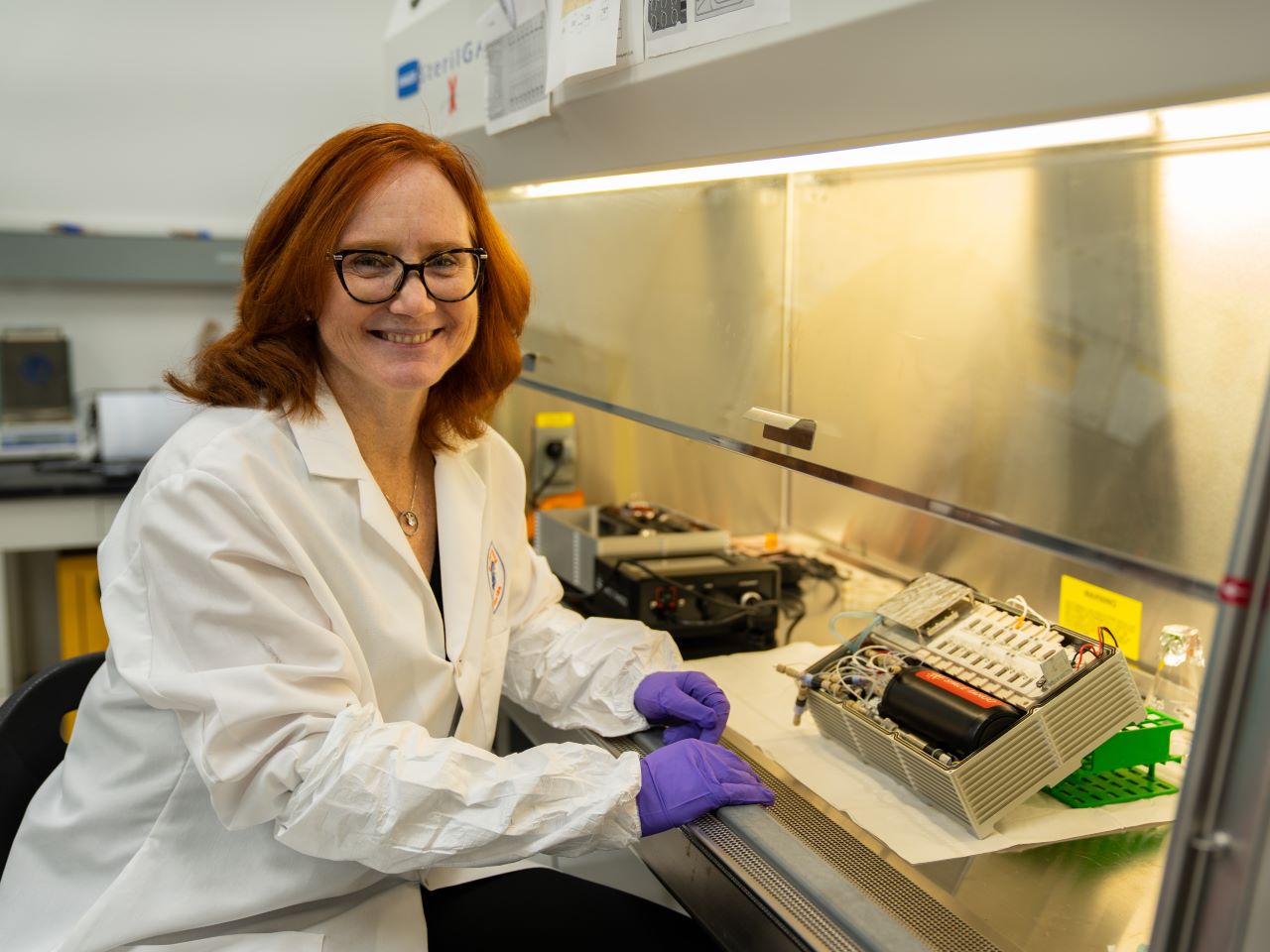
UF scientist studies muscle loss in space to benefit astronauts and patients on Earth
Astronauts traveling to Mars will face many challenges, but one of the most serious is muscle loss during long space missions. A new study led by University of Florida researcher Siobhan Malany, Ph.D., sheds light on how human biology changes in microgravity and could help protect astronaut health while also offering hope for patients with muscle-wasting diseases on Earth. Malany, an associate professor in the College of Pharmacy, a member of UF’s Astraeus Space Institute, and director of the in-space Biomanufacturing Innovation Hub, recently published findings showing how muscle cells adapt in space. Her team studied bioengineered three-dimensional muscle tissues derived from biopsy cells from both younger and older individuals and observed how they responded to electrical stimulation in microgravity. These micro-scale tissues called “tissue chips” were given nutrients and electric pulses autonomously in a miniature laboratory the size of a shoe box called a CubeLab.x. A camera system inside the box recorded the rate of muscle contraction. “This research is about more than just space,” Malany said. “By understanding how muscle tissue deteriorates much faster in microgravity, we can uncover new strategies to address muscle loss that occurs naturally with aging and with age-related diseases here on Earth.” Siobhan Malany studies the effects of microgravity on human muscle biology using an automated tissue chip system. View her profile here The study found that younger muscle tissue showed more pronounced changes in mitochondrial pathways — cellular systems that produce energy — than older tissue did when exposed to microgravity. Researchers also discovered that, on Earth, older muscle tissue responds less to electrical stimulation than younger tissue. But in space, the younger tissue showed a noticeable drop in its ability to contract, suggesting that younger muscle may experience a greater change when exposed to the space environment. These insights may help researchers design new treatments to protect muscles in astronauts during long missions, as well as develop therapies for people experiencing age-related muscle loss on Earth. The project was part of UF’s broader efforts to advance space biology. Through the Astraeus Space Institute, UF brings together experts across disciplines, from medicine and pharmacy to engineering and plant science, to address the unique challenges of space exploration. “UF researchers are helping lay the groundwork for humanity’s next giant leap,” Malany said. “It’s exciting to see our work contribute to both the health of astronauts and the lives of patients back home.” UF’s leadership in space biology is strengthened through collaboration with partners including the Kennedy Space Center Consortium and the Center for Science, Technology and Advanced Research in Space), both initiatives bringing together universities in Florida’s high-tech corridor, government agencies and industry leaders. Malany’s work also builds on long-term collaborations with AdventHealth, using donated tissue samples to model age-related muscle changes in space. Her team also works with SpaceTango, a NASA-certified aerospace company, to design the CubeLab that flew to the International Space Station on multiple SpaceX missions. Looking ahead, Malany and her team are developing new ways to study astronaut-derived cells, including both skeletal and heart muscle, generated from blood samples. These “avatars” could help researchers track changes before, during and after space missions, providing an unprecedented window into how microgravity affects the human body. “Now we can study cells from individual astronauts and see how they respond over time,” Malany said. “This helps us understand the risks of long-term spaceflight and also gives us a platform for testing potential treatments for muscle-wasting conditions on Earth.” By using tissue chips, small, bioengineered devices that mimic the structure and function of human organs, scientists in space can gather data more quickly and accurately than with traditional animal studies, potentially accelerating the discovery of therapies for aging-related muscle loss. Looking to know more about this amazing research or connect with Siobhan Malany - simply click on her icon now to arrange an interview today.


Nursing researcher receives over $500K in prestigious grants
For the first time in nearly 15 years, a faculty member from Augusta University’s College of Nursing has been awarded a grant from the National Institutes of Health. Blake McGee, PhD, has secured an R03 award of $176,331 from the Eunice Kennedy Shriver National Institute of Child Health and Human Development to study Medicaid’s expanded role in late postpartum maternal health. But he hasn’t stopped there as McGee is also part of the fifth cohort of Betty Irene Moore Fellows, a prestigious program for nurse leaders and innovators that has awarded CON half a million dollars to support his research project and leadership development. McGee, the prelicensure department chair and an associate professor, is collaborating with colleagues from other Georgia universities on both studies, which are occurring simultaneously. “I began my career as an ER nurse and have always wanted to ask bigger questions about the challenges facing patients and how we might best address them as a society,” said McGee, who was recently selected for publication in Blood Advances, the American Society of Hematology’s journal. “As nursing scientists, we are uniquely poised to ask questions about healthcare policy, specifically from the vantage point of the impact that policy choices have on patients and their health outcomes.” This century, the United States has seen rising maternal mortality rates with alarming racial disparities. Over half of these deaths occur in the postpartum period, with 23% occurring more than six weeks after delivery. Medicaid expansion covers pregnant women in households below 138% of the Federal poverty level through postpartum day 60, which has been associated with decreased mortality and reduced racial disparity in maternal death. At the time of grant submission, pregnancy Medicaid eligibility traditionally lapsed 60 days after delivery, leaving postpartum people vulnerable to disruptions in care. McGee’s work aims to identify changes in maternal health care use and health outcomes 60 days to 1 year after delivery that were associated with state Medicaid expansions (2007–19). The team will examine whether the effects of expansion vary by maternal race or ethnicity and will explore whether patient-reported health care access and quality mediate the relationships between expansion and outcomes. “My hope is that after the study we’ll have a better understanding of how health and health care use change for women in this crucial late postpartum period and how they may differ for people of different backgrounds,” said McGee. “Due to the sample design, findings will reliably inform optimal policy for postpartum coverage duration.” He expects this study to provide preliminary data for a future R01-funded study that directly examines the impact of extending the duration of postpartum Medicaid under the American Rescue Plan. As part of the Betty Irene Moore Fellowship, McGee is one of 15 fellows across the nation in a curriculum co-delivered by the UC Davis School of Nursing and Graduate School of Management. A project coordinator from AU’s School of Public Health will also assist with the fellowship project. McGee hopes to involve graduate research assistants or recent alumni as research associates on the team. Specifically, McGee will be studying the Georgia Pathways to Coverage Program, making him one of the only academic researchers in the nation funded to do so. “As a researcher, it is always a privilege to engage in topics that directly impact the current state of health care, and I’m honored to tackle projects that are so relevant to today’s health policy headlines,” he said. Georgia stands out among other states that are exploring an extension of Medicaid to low-income, working-age adults who demonstrate a monthly commitment of 80 hours to an employment-related activity. By studying the effects of this program, McGee predicts the findings will be highly relevant to anticipating the impact of recent Medicaid changes at the federal level and may indicate differences between Pathways participants and those who might qualify but remain uninsured. This focus could provide data that helps the state target enrollment efforts. The state’s own logic model predicts that the program will reduce hospitalizations, and McGee is eager to determine the program’s success. “Our findings should be helpful to the state to better understand those enrolling, what their experience with increased access to care has been and how their health has improved after receiving coverage,” McGee said.

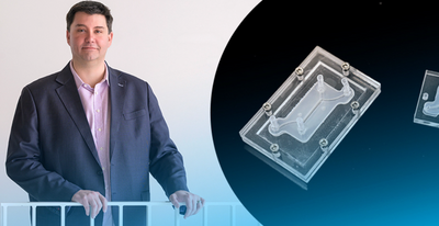
Taking discoveries to the real world for the benefit of human health
It takes about a decade and a lot of money to bring a new drug to market—between $1 billion to $2 billion, in fact. University of Delaware inventor Jason Gleghorn wants to change that. At UD, Gleghorn is developing leading-edge microfluidic tissue models. The devices are about the size of two postage stamps, and they offer a faster, less-expensive way to study disease and to develop pharmaceutical targets. These aren’t tools he wants to keep just for himself. No, Gleghorn wants to put the patented technology he’s developing in the hands of other experts, to advance clinical solutions in women’s health, maternal-fetal health and pre-term birth. His work also has the potential to improve understanding of drug transport in the female reproductive tract, placenta, lung and lymph nodes. Gleghorn, an associate professor of biomedical engineering, was named to the first cohort of Innovation Ambassadors at UD, as part of the University’s effort to foster and support an innovation culture on campus. Below, he shares some of what he’s learned about translating research to society. Q: What is the problem that you are trying to address? Gleghorn: A lot of disease has to do with disorganization in the body’s normal tissue structure. My lab makes microfluidic tissue models, called organ-on-a-chip models, that have super-tiny channels about the thickness of a human hair, where we can introduce very small amounts of liquid, including cells, to represent an organ in the human body. This can help us study and understand the mechanism of how things work in the body (the biology) or help us do things like drug screening to test therapeutic compounds for treating disease. And while these little microfluidic devices can do promising things, the infrastructure required to make the system work often restricts their use to high-end labs. We want to democratize the techniques and technology so that nonexperts can use it. To achieve this, we changed the way we make these devices, so that they are compatible with standard manufacturing, which means we can scale them and create them much easier. Gleghorn: One of the problems with drug screening, in general, is that animal model studies don’t always represent human biology. So, when we’re using animal models to test new drugs — which have been the best tool we have available — the results are not always apples to apples. Fundamentally, our microfluidic devices can model what happens in humans … we can plug in the relevant human components to understand how the mechanism is working and then ask questions about what drives those processes and identify targets for therapies to prevent the dysfunction. Q: What is innovative about this device? Gleghorn: The innovation part is this modularity — no one makes these devices this way. The science happens on the tiny tissue model insert, which is sandwiched between two pieces of clear acrylic. This allows us to watch what’s happening on the tissue model insert in real time. Meanwhile, the outer shell’s clamshell design provides flexibility: if we’re studying lung tissue and we want to study the female reproductive tract, all we do is unscrew the outer shell and insert the proper tissue model that mimics the female reproductive tract and we’re off. We’ve done a lot of the engineering to make it very simple to operate and use, and adaptable to common lab tools that everyone has, to eliminate the need for financial investment in things like specialized clean rooms, incubators and pumps, etc., so the technology can be useful in regular labs or easily deployable to far-flung locations or countries. With a laser cutter and $500 worth of equipment, you could conceivably mass manufacture these things for maternal medicine in Africa, for example. Democratizing the technology so it is compatible and useful for even an inexperienced user aligns with the mission of my lab, which focuses on scaling the science and the innovation faster, instead of only a few specialized labs being a bottleneck to uncovering new mechanisms of disease and the development of therapies. We patented this modularity, the way to build these tiny microfluidic devices and the simplicity of how it's used as a tool set, through UD’s Office of Economic Innovation and Partnerships (OEIP). Q: How have you translated this work so far? Gleghorn: To date, we've taken this microfluidic system to nine different research labs across seven countries and four continents — including the United States, the United Kingdom, Australia, France, Belgium and South Africa. These labs are using our technology to study problems in women’s health and collecting data with it. We’re developing boot camps where researchers can come for two or three days to the University of Delaware, where we teach them how to use this device and they take some back with them. From a basic science perspective, there is high enthusiasm for the power of what it can tell you and its ease of use. As engineers, we think it's pretty cool that many other people are using our innovations for new discoveries. Q: What support and guidance have you received from the UD innovation ecosystem? Gleghorn: To do any of this work, you need partners that have various expertise and backgrounds. UD’s Office of Economic Innovation and Partnerships has built a strong team of professionals with expertise in different areas, such as how do you license or take something to patent, how do you make connections with the business community? OEIP is home to Delaware’s Small Business Development Center, which can help you think about business visibility in terms of startups. Horn Entrepreneurship has built out impressive programs for teaching students and faculty to think entrepreneurially and build mentor networks, while programs like the Institute for Engineering Driven Health and the NSF Accelerating Research Translation at UD provide gap funding to be able to do product development and to take the work from basic prototype to something that is more marketable. More broadly in Delaware is the Small Business Administration, the Delaware Innovation Space and regional grant programs and small accelerators to help Delaware innovators. Q: How have students in your lab benefited from engaging in innovation? Gleghorn: Undergraduate students in my lab have made hundreds of these devices at scale. We basically built a little manufacturing facility, so we have ways to sterilize them, track batches, etc. We call it “the foundry.” In other work, graduate students are engineering different components or working on specific system designs for various studies. The students see collaborators use these devices to discover new science and new discoveries. That's very rewarding as an engineer. Additionally, my lab focuses on building solutions that are useful in the clinic and commercially viable. As a result, we've had two grad students spin out companies related to the work we've been doing in the lab. Q: How has research translation positively impacted your work? Gleghorn: I started down this road maybe five years ago, seriously trying to think about how to translate our research findings. Being an entrepreneur, translating technology — it's a very different way to think about your work. And so that framework has really permeated most of the research that I do now and changed the way I think about problems. It has opened new opportunities for collaboration and for alternate sources of funding with companies. This has value in terms of taking the research that you're doing fundamentally and creating a measurable impact in the community, but it also diversifies your funding streams to work on important problems. And different viewpoints help you look at the work you do in new ways, challenging you to define the value proposition, the impact of your work.

ChristianaCare Breaks Ground on New Middletown Health Center
ChristianaCare today broke ground on its new Health Center at Middletown, marking a major milestone in bringing expanded, affordable and exceptional care to families in southern New Castle County and northern Kent County. The center is expected to open in spring 2027. The $92.3 million project reflects a deep investment in the health and vitality of the region and is part of ChristianaCare’s larger plan, announced in July, to invest more than $865 million in Delaware over the next three years. The 87,000-square-foot Health Center will rise at 621 Middletown Odessa Road, next to ChristianaCare’s existing freestanding emergency department. Designed as a modern, multidisciplinary hub, the facility will expand access to comprehensive services and create more than 70 new full-time jobs, boosting both community health and the local economy. “Today we take an exciting step forward for Delaware, as part of ChristianaCare’s $865 million investment to expand access and strengthen health across our state,” said Janice E. Nevin, M.D., MPH, President and CEO of ChristianaCare. “This new health center is a promise to Delawareans: that they will have access to exceptional care close to home, delivered with love and excellence. More than a building, it represents our vision for healthier communities, our deep commitment to those we serve, and a future where every neighbor can thrive.” A Holistic, Patient-Centered Experience The ChristianaCare Health Center at Middletown will bring together a wide range of services in one convenient location, including: Primary and specialty care. Women’s health, behavioral health, oncology, cardiovascular care, pediatrics, neurology, imaging, diagnostics and lab testing. Hybrid exam rooms with interactive digital tools that allow family members to join virtually. Calming waiting areas with sensory-sensitive design features, plus friendly floor ambassadors to help patients navigate the building. Healing environments that include walking trails and abundant natural light. “We are designing care around people, not around appointments or buildings,” said Pauline Corso, president of Ambulatory Network Continuity and Growth at ChristianaCare. “From easy parking to advanced care coordination, every detail of this new center is aimed at making health care more welcoming, more connected and more human.” A Community Partnership ChristianaCare has been part of the Middletown community since 2009, when it first acquired the land that is now home to the freestanding emergency department. Last year, that facility provided care for more than 32,000 patient visits. “This groundbreaking is a proud moment for our town,” said Ken Branner, mayor of Middletown. “ChristianaCare has been a trusted partner for many years, and this new facility shows a lasting commitment to our residents. It will bring top-quality care closer to home and create good jobs right here in our community.”