Experts Matter. Find Yours.
Connect for media, speaking, professional opportunities & more.
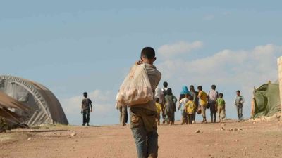
Violence alters human genomes for generations, researchers discover
In February of 1982, the Syrian government besieged the city of Hama, killing tens of thousands of its own citizens in sectarian violence. Four decades later, rebels used the memory of the massacre to help inspire the toppling of the Assad family that had overseen the operation. But there is another lasting effect of the attack, hidden deep in the genes of Syrian families. The grandchildren of women who were pregnant during the siege — grandchildren who never experienced such violence themselves — nonetheless bear marks of it in their genomes. Passed down through their mothers, this genetic imprint offers the first human evidence of a phenomenon previously documented only in animal models. The genetic transmission of stress across multiple generations. “The idea that trauma and violence can have repercussions into future generations should help people be more empathetic, help policymakers pay more attention to the problem of violence,” said Connie Mulligan, Ph.D., a professor of Anthropology and the Genetics Institute at the University of Florida and co-senior author of the new study. “It could even help explain some of the seemingly unbreakable intergenerational cycles of abuse and poverty and trauma that we see around the world, including in the U.S.” While our genes are not changed by life experiences, they can be tuned through a system known as epigenetics. In response to stress or other events, our cells can add small chemical flags to genes that may quiet them down or alter their behavior. These changes may help us adapt to stressful environments, although the effects aren’t well understood. It is these tell-tale chemical flags that Mulligan and her team were looking for in the genes of Syrian families. While lab experiments have shown that animals can pass along epigenetic signatures of stress to future generations, proving the same in people has been nearly impossible. “Resilience and perseverance is quite possibly a uniquely human trait.” —Connie Mulligan Mulligan worked with Rana Dajani, Ph.D., a molecular biologist at Hashemite University in Jordan and co-senior author, as well as anthropologist Catherine Panter-Brick, Ph.D., of Yale University, to conduct the unique study. Dajani envisioned the research project; because of her intimate knowledge of the Syrian population and its tragic history, she designed the study to cover three generations of Syrian refugees to Jordan. Some families had lived through the Hama attack before fleeing to Jordan. Other families avoided Hama, but lived through the recent civil war against the Assad regime. The team collected samples from grandmothers and mothers who were pregnant during the two conflicts, as well as from their children. This study design meant there were grandmothers, mothers and children who had each experienced violence at different stages of development. A third group of families had immigrated to Jordan before 1980, avoiding the decades of violence in Syria. These early immigrants served as a crucial control to compare to the families who had experienced the stress of civil war. Study coauthor Dima Hamadmad, a Syrian researcher and the daughter of refugees, led the search for families that met the study criteria and collected cheek swabs from 138 people across 48 families. "The participants took part in the research out of love for their children and concern for future generations,” she said. “But more than that, they wanted their stories of trauma to be heard and acknowledged.” Back in Florida, Mulligan’s lab scanned the DNA for epigenetic modifications and looked for any relationship with the families’ experience of violence. In the grandchildren of Hama survivors, the researchers discovered 14 areas in the genome that had been modified in response to the violence their grandmothers experienced. These 14 modifications demonstrate that stress-induced epigenetic changes may indeed appear in future generations in humans, just as they can in animals. The study also uncovered 21 epigenetic sites in the genomes of people who had directly experienced violence in Syria. In a third finding, the researchers reported that people exposed to violence while in their mothers’ wombs showed evidence of accelerated epigenetic aging, a type of biological aging that may be associated with susceptibility to age-related diseases. Most of these epigenetic changes showed the same pattern after exposure to violence, suggesting a kind of common epigenetic response to stress – one that can not only affect people directly exposed to stress, but also future generations. “We think our work is relevant to many forms of violence, not just refugees. Domestic violence, sexual violence, gun violence: all the different kinds of violence we have in the U.S,” said Mulligan. “We should study the effects of violence. We should take it more seriously.” It’s not clear what, if any, effect these epigenetic changes have in the lives of people carrying them inside their genomes. But some studies have found a link between stress-induced epigenetic changes and diseases like diabetes. One famous study of Dutch survivors of famine during World War II suggested that their offspring carried epigenetic changes that increased their odds of being overweight later in life. While many of these modifications likely have no effect, It’s possible that some have functional effects that can affect our health, Mulligan said. The researchers published their findings, which were supported by the National Science Foundation, Feb. 27 in the journal Scientific Reports. While carefully searching for evidence of the lasting effects of war and trauma stamped into our genomes, Mulligan and her collaborators were also struck by the perseverance of the families they worked with. Their story was much bigger than merely surviving war, Mulligan said. “In the midst of all this violence we can still celebrate their extraordinary resilience. They have persevered,” Mulligan said. “That resilience and perseverance is quite possibly a uniquely human trait.”

UF expert answers questions about local risk of bird flu
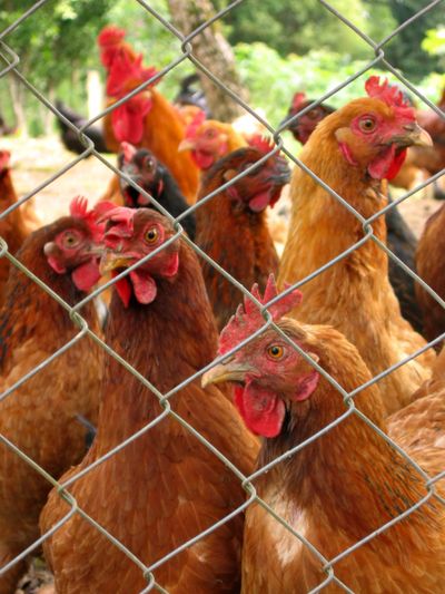
Consumers may have noticed the rising price of eggs and even some shortages at grocery stores lately due to H5N1 avian influenza, but as cases of human and animal infections continue to rise, how concerned should you be about the virus? Benjamin Anderson, Ph.D., an assistant professor in the University of Florida College of Public Health and Health Professions’ Department of Environmental and Global Health and lead for UF’s Emerging Pathogens Institute bird flu response team answers some questions about the risk of infection to humans and animals from bird flu and how to protect yourself. Who is at risk? Currently, the Centers for Disease Control and Prevention says the risk to humans is low. “That is correct on an overall level,” Anderson said. There is no human-to-human transmission right now. Anderson said that while there have been an “alarming” number of human cases, the number of infections is still fewer than 100. Of those, most have resulted in mild illness and were in people who had direct exposure to infected animals. So far, there has been only one death attributed to the current outbreak of H5N1, known more commonly as bird flu – a man in Louisiana who was infected by a backyard flock. “We do have a lot of people who keep chickens,” Anderson said. “Because of the situation in Louisiana, this has, I think, piqued the concern even more so among folks who might have backyard poultry to recognize that is a potential pathway for transmission.” If you see a dead chicken, do not touch it or try to investigate yourself. Instead, report it to the Florida Department of Agriculture and Consumer Services. Report dead wildlife, including migratory birds, to the Florida Fish and Wildlife Commission. Anderson said while the risk to the public is currently low, the future risk, including human-to-human transmission that could result in a pandemic, is still uncertain. Right now, he said, “Unless you’re handling poultry or working with or near dairy cattle, where bird flu outbreaks have been ongoing, your risk is relatively low.” Can I catch the virus from my backyard bird feeder? Gainesville is on a major flyway for migratory birds – a draw for birdwatching enthusiasts, particularly in the winter. This may be how bird flu has made its way into backyard flocks, since infected migratory birds shed the virus in their waste. So far, there is no data that suggests bird feeders could pose a significant transmission risk to people, and no reported human cases of bird flu have been traced to feeders. “However, when I say there’s no data, that means there’s no data. It hasn’t been investigated,” Anderson said. Waterfowl such as ducks and geese are more likely to carry the virus than songbirds. But if backyard birdwatchers are concerned, he said, take precautions such as wearing gloves and disinfecting bird feeders. And whether there’s an elevated risk of bird flu or not, always take care to avoid touching bird feces, which can contain salmonella. “Using some common sense, good hygiene practices, is going to be an effective way of protecting yourself,” he said. Are my outdoor cats in danger? Outdoor cats are susceptible to bird flu through exposure to dairy cattle, wild birds and contaminated raw milk. There is also new evidence of some sources of raw cat food being contaminated with H5N1. H5N1 causes severe infection in cats, with neurological symptoms that could mimic rabies. Infected cats may be disoriented, lethargic or disinterested in food. Florida residents can contact the Florida Health Department if they notice these symptoms in their pet. “If you see something unusual, seeking out professional care for that animal is an important thing to do,” Anderson said. As of now, it’s unclear whether cats can transmit the virus to humans, but as it adapts, transmission to other species could become easier and more widespread. What about eggs and poultry from the farmers market? Florida state statutes require sellers to register as a food supplier and meet certain criteria for food handling safety. But the regulations can be unclear to some small, local egg and poultry producers, and others operate under the radar. “I wouldn’t say that if you go to a farmers market, it’s a guarantee that the products you’re buying are produced under the proper regulations,” Anderson said, but the regulations themselves can be unclear. Some things you can do to keep yourself safe are asking the vendor if they’re registered and permitted with the state’s agriculture agency and checking that the products are labeled. Per the statutes, eggs must be refrigerated at all times between packaging and sale to the consumer. And definitely steer clear of raw milk, which has been tied to several human and animal H5N1 infections and always carries a risk of salmonella. “Don’t drink it, and don’t give it to your animals,” Anderson said. Is there a vaccine? How else can I protect myself? There is a vaccine for bird flu. While it isn’t currently being administered to humans in the U.S., Anderson said some agriculture workers in Europe have received it. “There is a potential justification for starting to release some of the stock of the H5N1-specific vaccine,” but it would come with tradeoffs, he said, such as maintaining stockpiles and keeping the vaccine matched well to an evolving virus. In his opinion, though, it makes sense to start the process now, both to protect workers who are already at higher risk of contracting the virus, and to begin collecting data on how well the vaccine is working. The idea that the U.S. should hold off on releasing a vaccine until bird flu becomes a pandemic is contrary to protecting public health, he said, adding that the tipping point for him was seeing the virus start to show up in backyard poultry. “That’s the rationale that I base my opinion off of,” Anderson said.

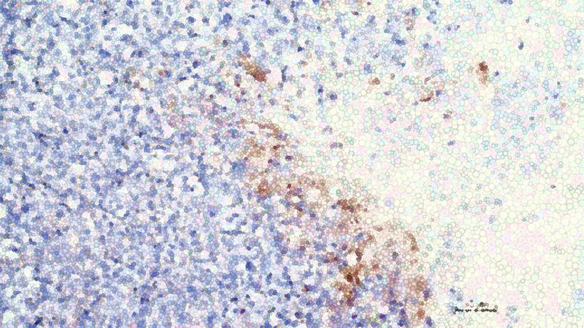
In a major step forward for cancer care, researchers at ChristianaCare’s Gene Editing Institute have shown that disabling the NRF2 gene with CRISPR technology can reverse chemotherapy resistance in lung cancer. The approach restores drug sensitivity and slows tumor growth. The findings were published Nov. 13, 2025 in the online edition of Molecular Therapy Oncology. This breakthrough stems from more than a decade of research by the Gene Editing Institute into the NRF2 gene, a known driver of treatment resistance. The results were consistent across multiple in vitro studies using human lung cancer cell lines and in vivo animal models. “We’ve seen compelling evidence at every stage of research,” said Kelly Banas, Ph.D., lead author of the study and associate director of research at the Gene Editing Institute. “It’s a strong foundation for taking the next step toward clinical trials.” Potential Beyond Lung Cancer The study focused on lung squamous cell carcinoma, an aggressive and common form of non-small cell lung cancer (NSCLC) that accounts for 20% to 30% of all lung cancer cases, according to the American Cancer Society. It’s estimated that over 190,000 people in the U.S. will be diagnosed in 2025. While the research centered on this cancer type, the implications are broader. Overactive NRF2 contributes to chemotherapy resistance in several solid tumors, including liver, esophageal and head and neck cancers. The results suggest a CRISPR-based strategy targeting NRF2 could help resensitize a wide range of treatment-resistant tumors to standard chemotherapy. “This is a significant step toward overcoming one of the biggest challenges in cancer therapy — drug resistance,” Banas said. “By targeting a key transcription factor that drives resistance, we’ve shown that gene editing can re-sensitize tumors to standard treatment. We’re hopeful that in clinical trials and beyond, this is what will allow chemotherapy to improve outcomes for patients and could enable them to remain healthier during the entirety of their treatment regimen.” Targeting a Master Switch for Resistance The research zeroed in on a tumor-specific mutation, R34G, in the NRF2 gene, which acts as a master regulator of cellular stress responses. When overactive, NRF2 helps cancer cells withstand chemotherapy. Using CRISPR/Cas9, the team engineered lung cancer cells with the R34G mutation and successfully knocked out NRF2. This restored sensitivity to chemotherapy drugs such as carboplatin and paclitaxel. In animal models, tumors directly treated with CRISPR to knockout NRF2 grew more slowly and responded better to treatment. “This work brings transformational change to how we think about treating resistant cancers,” said Eric Kmiec, Ph.D., senior author of the study and executive director of the Gene Editing Institute. “Instead of developing entirely new drugs, we are using gene editing to make existing ones effective again.” Editing Reaches Threshold Levels One of the most promising discoveries was that disrupting NRF2 in just 20% to 40% of tumor cells, was enough to improve the response to chemotherapy and shrink tumors. This insight is particularly relevant for clinical use, where editing every cancer cell may not be feasible. To test therapy in mice, the researchers used lipid nanoparticles (LNPs), a non-viral method with high efficiency and low risk of unintended, off-target effects. Sequencing confirmed that the edits were highly specific to the mutated NRF2 gene, with minimal unintended changes elsewhere in the genome. “The power of this CRISPR therapy lies in its precision. It’s like an arrow that hits only the bullseye,” said Banas. “This level of specificity with minimal unanticipated genomic side effects offers real hope for the cancer patients who could one day receive this treatment.”


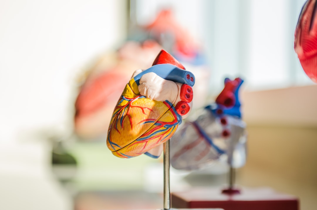
Heart valve developed at UC Irvine shines in early-stage preclinical testing
UC Irvine researchers designed and developed a minimally invasive replacement pulmonary heart valve. Created for pediatric patients, the device can be expanded as children grow, eliminating the need for multiple surgeries. The team successfully conducted laboratory and early-stage animal feasibility testing of the implant, crucial steps toward approval for human use. Irvine, Calif., June 23, 2025 — Researchers at the University of California, Irvine have successfully performed preclinical laboratory testing of a replacement heart valve intended for toddlers and young children with congenital cardiac defects, a key step toward obtaining approval for human use. The results of their study were published recently in the Journal of the American Heart Association. The management of patients with congenital heart disease who require surgical pulmonary valve replacement typically occurs between the ages of 2 and 10. To be eligible for a minimally invasive transcatheter pulmonary valve procedure, patients currently must weigh at least 45 pounds. For children to receive minimally invasive treatment, they must be large enough so that their veins can accommodate the size of a crimped replacement valve. The Iris Valve designed and developed by the UC Irvine team can be implanted in children weighing as little as 17 to 22 pounds and gradually expanded to an adult diameter as they grow. Research and development of the Iris Valve has been supported by the Eunice Kennedy Shriver National Institute of Child Health and Human Development; the National Heart, Lung, and Blood Institute; and the National Science Foundation. This funding has enabled benchtop fracture testing, which demonstrated the valve’s ability to be crimped down to a 3-millimeter diameter for transcatheter delivery and subsequently enlarged to 20 millimeters without damage, as well as six-month animal studies that confirmed successful device integration within the pulmonary valve annulus, showing valve integrity and a favorable tissue response. “We are pleased to see the Iris Valve performing as we expected in laboratory bench tests and as implants in Yucatan mini pigs, a crucial measure of the device’s feasibility,” said lead author Arash Kheradvar, UC Irvine professor of biomedical engineering. “This work represents the result of longstanding collaboration between our team at UC Irvine and Dr. Michael Recto at Children’s Hospital of Orange County built over several years of joint research and development.” Congenital heart defects affect about 1 percent of children born in the United States and Europe, with over 1 million cases in the U.S. alone. These conditions often necessitate surgical interventions early in life, with additional procedures required to address a leaky pulmonary valve and prevent right ventricular failure as children grow. The Iris Valve can be implanted via a minimally invasive catheter through the patient’s femoral vein. The Kheradvar group employed origami folding techniques to compress the device into a 12-French transcatheter system, reducing its diameter to no more than 3 millimeters. Over time, the valve can be balloon-expanded up to its full 20-millimeter diameter. This implantation method, along with the ability to begin treatment earlier in very young patients, helps mitigate the risk of complications from delayed care and reduces the need for multiple surgeries in this vulnerable population. “Once the Iris Valve comes to fruition, it will save hundreds of children at least one operation – if not two – throughout the course of their lives,” said Recto, an interventional pediatric cardiologist at CHOC who’s also a clinical professor of pediatrics at UC Irvine. “It will save them from having to undergo surgical pulmonary valve placement, as the Iris Valve is delivered via a small catheter in the vein and can be serially dilated to an adult diameter and also facilitate the future placement of larger transcatheter pulmonary valves – with sizes greater than 20 millimeters, like the Melody, Harmony and Sapien devices – if needed.” Kheradvar said that the next phase of preclinical testing of the Iris Valve is funded by the Brett Boyer Foundation, which is committed to supporting research into treatments for congenital heart disease. “We are actively engaged with the U.S. Food and Drug Administration to define and carry out the required experiments and documentation for first-in-human authorization of the Iris Valve,” Kheradvar said. “Our team is urgently advancing the Iris Valve through preclinical studies to enable its clearance for first-in-human use. This is a critical step toward providing toddlers – who currently have no viable minimally invasive treatment until they reach the 45-pound threshold – with a much-needed option.” First co-author Nnaoma Agwu, a biomedical engineering Ph.D. candidate at UC Irvine, said: “The development of the Iris Valve required a strong and knowledgeable team that understood the clinical and mechanical design requirements. This accomplishment would not have been possible without the collaboration of talented clinicians, veterinarians and engineers. With this milestone reached, we are rigorously advancing the Iris Valve’s development, setting our sights on human clinical trials.” Joining Kheradvar, Recto and Agwu as co-authors of the article in Journal of the American Heart Association were Daryl Chau, a recent UC Irvine master’s graduate; Gregory Kelley and Tanya Burney, both research specialists at UC Irvine, with Burney also affiliated with the Beckman Laser Institute; Ekaterina Perminov, a clinical veterinarian with UC Irvine’s University Laboratory Animal Resources; and Christopher Alcantara, a radiology technician at CHOC. About UC Irvine’s Brilliant Future campaign: Publicly launched on Oct. 4, 2019, the Brilliant Future campaign aims to raise awareness and support for the university. By engaging 75,000 alumni and garnering $2 billion in philanthropic investment, UC Irvine seeks to reach new heights of excellence in student success, health and wellness, research and more. The Samueli School of Engineering plays a vital role in the success of the campaign. Learn more by visiting https://brilliantfuture.UC Irvine.edu/the-henry-samueli-school-of-engineering About the University of California, Irvine: Founded in 1965, UC Irvine is a member of the prestigious Association of American Universities and is ranked among the nation’s top 10 public universities by U.S. News & World Report. The campus has produced five Nobel laureates and is known for its academic achievement, premier research, innovation and anteater mascot. Led by Chancellor Howard Gillman, UC Irvine has more than 36,000 students and offers 224 degree programs. It’s located in one of the world’s safest and most economically vibrant communities and is Orange County’s second-largest employer, contributing $7 billion annually to the local economy and $8 billion statewide. For more on UC Irvine, visit www.uci.edu. Media access: Radio programs/stations may, for a fee, use an on-campus studio with a Comrex IP audio codec to interview UC Irvine faculty and experts, subject to availability and university approval. For more UC Irvine news, visit news.uci.edu. Additional resources for journalists may be found at https://news.uci.edu/media-resources.

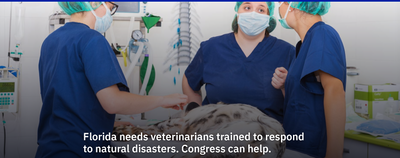
Florida needs veterinarians trained to respond to natural disasters. Congress can help.
When Hurricane Helene struck, we were on the frontlines in Florida’s Big Bend region, racing against time to support the Humane Society as they rescued animals displaced by the most powerful storm ever to hit this part of the state. Two weeks later, we were back in action, facing the devastating flooding from Hurricane Milton. These back-to-back disasters showcased the urgency and critical need for emergency-response veterinarians who can act fast to save lives. We lead one of the nation’s only three emergency veterinary response teams — the University of Florida Veterinary Emergency Treatment Service (UF VETS). Founded after the 2004 hurricane season and operating under the UF College of Veterinary Medicine, the UF VETS program hosts two distinct, yet complementary, branches: a medical response unit for disaster-affected animals and an animal technical rescue branch, which manages complex operations like overturned livestock trailers. Larry Garcia specializes in veterinary disaster preparedness and response, animal technical rescue/training and shelter medicine operations. View his profile here Our team is on call whenever disaster strikes, working alongside local and state veterinary organizations, animal rescues and law enforcement to save animals in crisis. But here’s the problem: Without a nationwide system for coordinating these efforts, it’s often chaotic, and animals suffer because of it. Now Congress has a golden opportunity to change that. As they return to Washington, they have the chance to make a game-changing impact by including funding in the final FY 2025 Homeland Security Appropriations bill to create a nationwide network of veterinary emergency teams. This funding could revolutionize how the U.S. handles animal care during national disasters — and it needs to happen, fast. Read more ... Looking to know more about this important topic or connect with Lawrence Garcia - simply click on his icon now to arrange an interview today.

3 Things A Climate Scientist Learned From Jane Goodall
In a recent Forbes article, Marshall Shepherd reflects on three key lessons he has drawn from the life and work of Dr. Jane Goodall. Shepherd frames Goodall’s legacy—spanning primatology, conservation, and public engagement—as deeply instructive for climate scientists and environmental advocates. He argues that her methods and mindset have more to teach than simply how to observe nature; they speak to how we engage with the world. First, Shepherd highlights immersion: Goodall’s decades of patient observation in the Tanzanian forests demonstrates the power of being physically—and emotionally—present to truly learn from ecosystems. For Shepherd, climate science must go beyond remote data collection: getting into the field and understanding local realities matters. Second, he emphasizes patience. Goodall’s willingness to wait, sometimes for years, for breakthroughs in understanding primate behavior offers a lesson for climate researchers, whose progress may unfold over decades. Third, he admires her tenacity—a commitment sustained over a lifetime, even under adversity. Shepherd suggests that tackling climate change requires that same kind of enduring resolve, especially when public attention or funding waxes and wanes. Through these reflections, Shepherd presents Goodall not just as an icon of conservation but as a model for scientific humility and perseverance. He invites readers to see the parallels between animal behavior research and climate work—and to adopt practices of listening, patience, and resolve in confronting our planet’s changing trajectory. Dr. J. Marshall Shepherd is a leading international weather-climate expert and is the Georgia Athletic Association Distinguished Professor of Geography and Atmospheric Sciences at the University of Georgia. Dr. Shepherd was the 2013 President of American Meteorological Society (AMS), the nation’s largest and oldest professional/science society in the atmospheric and related sciences. View his profile here Dr. J. Marshall Shepherd is a leading international weather-climate expert and is the Georgia Athletic Association Distinguished Professor of Geography and Atmospheric Sciences at the University of Georgia. He's available to speak with the media about this topic - simply click on his icon now to arrange an interview today.

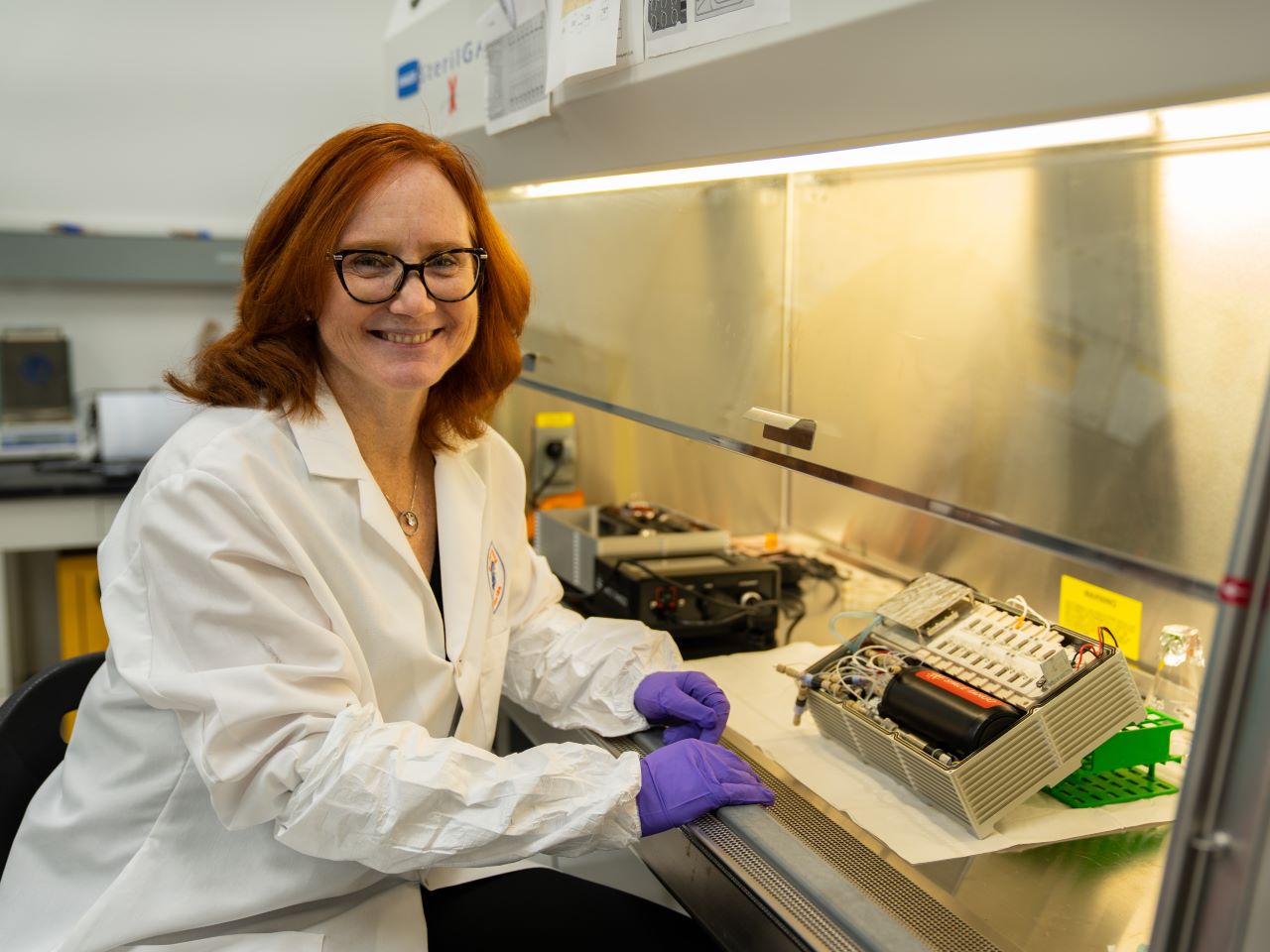
UF scientist studies muscle loss in space to benefit astronauts and patients on Earth
Astronauts traveling to Mars will face many challenges, but one of the most serious is muscle loss during long space missions. A new study led by University of Florida researcher Siobhan Malany, Ph.D., sheds light on how human biology changes in microgravity and could help protect astronaut health while also offering hope for patients with muscle-wasting diseases on Earth. Malany, an associate professor in the College of Pharmacy, a member of UF’s Astraeus Space Institute, and director of the in-space Biomanufacturing Innovation Hub, recently published findings showing how muscle cells adapt in space. Her team studied bioengineered three-dimensional muscle tissues derived from biopsy cells from both younger and older individuals and observed how they responded to electrical stimulation in microgravity. These micro-scale tissues called “tissue chips” were given nutrients and electric pulses autonomously in a miniature laboratory the size of a shoe box called a CubeLab.x. A camera system inside the box recorded the rate of muscle contraction. “This research is about more than just space,” Malany said. “By understanding how muscle tissue deteriorates much faster in microgravity, we can uncover new strategies to address muscle loss that occurs naturally with aging and with age-related diseases here on Earth.” Siobhan Malany studies the effects of microgravity on human muscle biology using an automated tissue chip system. View her profile here The study found that younger muscle tissue showed more pronounced changes in mitochondrial pathways — cellular systems that produce energy — than older tissue did when exposed to microgravity. Researchers also discovered that, on Earth, older muscle tissue responds less to electrical stimulation than younger tissue. But in space, the younger tissue showed a noticeable drop in its ability to contract, suggesting that younger muscle may experience a greater change when exposed to the space environment. These insights may help researchers design new treatments to protect muscles in astronauts during long missions, as well as develop therapies for people experiencing age-related muscle loss on Earth. The project was part of UF’s broader efforts to advance space biology. Through the Astraeus Space Institute, UF brings together experts across disciplines, from medicine and pharmacy to engineering and plant science, to address the unique challenges of space exploration. “UF researchers are helping lay the groundwork for humanity’s next giant leap,” Malany said. “It’s exciting to see our work contribute to both the health of astronauts and the lives of patients back home.” UF’s leadership in space biology is strengthened through collaboration with partners including the Kennedy Space Center Consortium and the Center for Science, Technology and Advanced Research in Space), both initiatives bringing together universities in Florida’s high-tech corridor, government agencies and industry leaders. Malany’s work also builds on long-term collaborations with AdventHealth, using donated tissue samples to model age-related muscle changes in space. Her team also works with SpaceTango, a NASA-certified aerospace company, to design the CubeLab that flew to the International Space Station on multiple SpaceX missions. Looking ahead, Malany and her team are developing new ways to study astronaut-derived cells, including both skeletal and heart muscle, generated from blood samples. These “avatars” could help researchers track changes before, during and after space missions, providing an unprecedented window into how microgravity affects the human body. “Now we can study cells from individual astronauts and see how they respond over time,” Malany said. “This helps us understand the risks of long-term spaceflight and also gives us a platform for testing potential treatments for muscle-wasting conditions on Earth.” By using tissue chips, small, bioengineered devices that mimic the structure and function of human organs, scientists in space can gather data more quickly and accurately than with traditional animal studies, potentially accelerating the discovery of therapies for aging-related muscle loss. Looking to know more about this amazing research or connect with Siobhan Malany - simply click on her icon now to arrange an interview today.

'Brain-on-a-chip': Engineering tomorrow’s breakthroughs today
A “brain-on-a-chip” technology might sound like science fiction, but it’s real-world hope. James McGrath, a biomedical engineer at the University of Rochester, leads a team that develops micro-scale tissue chips to study diseases in lieu of conducting animal experiments. The team’s “brain-on-a-chip” model replicates the blood-brain barrier — the critical membrane separating the brain from the bloodstream — to mimic how the barrier functions under healthy conditions and the duress of infections, toxins, and immune responses that can weaken it. Recent findings from McGrath’s team show how systemic inflammation, such as that caused by sepsis, can compromise the barrier and harm brain cells. The researchers also demonstrated how pericytes — supportive vascular cells — can help repair barrier damage, an insight that could guide new therapies for Alzheimer’s and Parkinson’s. The research culminated in a pair of recent studies published in Advanced Science and Materials Today Bio. “We hope that by building these tissue models in chip format, we can arrange many brain models in a high-density array to screen candidates for neuroprotective drugs and develop brain models with diverse genetic backgrounds,” McGrath says. McGrath aims to transform how scientists test drugs and predict neurological side effects before they occur — helping rewrite how we study, and one day safeguard, the brain. Contact McGrath by clicking on his profile

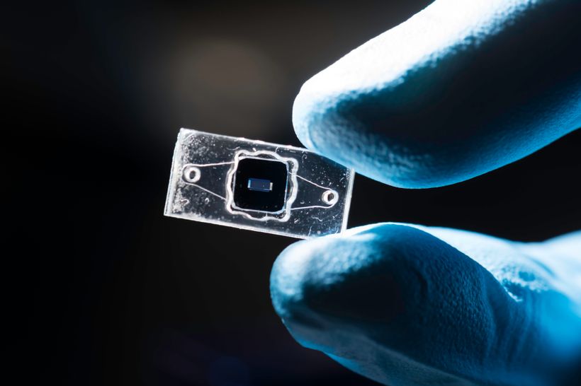
Taking discoveries to the real world for the benefit of human health
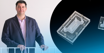
It takes about a decade and a lot of money to bring a new drug to market—between $1 billion to $2 billion, in fact. University of Delaware inventor Jason Gleghorn wants to change that. At UD, Gleghorn is developing leading-edge microfluidic tissue models. The devices are about the size of two postage stamps, and they offer a faster, less-expensive way to study disease and to develop pharmaceutical targets. These aren’t tools he wants to keep just for himself. No, Gleghorn wants to put the patented technology he’s developing in the hands of other experts, to advance clinical solutions in women’s health, maternal-fetal health and pre-term birth. His work also has the potential to improve understanding of drug transport in the female reproductive tract, placenta, lung and lymph nodes. Gleghorn, an associate professor of biomedical engineering, was named to the first cohort of Innovation Ambassadors at UD, as part of the University’s effort to foster and support an innovation culture on campus. Below, he shares some of what he’s learned about translating research to society. Q: What is the problem that you are trying to address? Gleghorn: A lot of disease has to do with disorganization in the body’s normal tissue structure. My lab makes microfluidic tissue models, called organ-on-a-chip models, that have super-tiny channels about the thickness of a human hair, where we can introduce very small amounts of liquid, including cells, to represent an organ in the human body. This can help us study and understand the mechanism of how things work in the body (the biology) or help us do things like drug screening to test therapeutic compounds for treating disease. And while these little microfluidic devices can do promising things, the infrastructure required to make the system work often restricts their use to high-end labs. We want to democratize the techniques and technology so that nonexperts can use it. To achieve this, we changed the way we make these devices, so that they are compatible with standard manufacturing, which means we can scale them and create them much easier. Gleghorn: One of the problems with drug screening, in general, is that animal model studies don’t always represent human biology. So, when we’re using animal models to test new drugs — which have been the best tool we have available — the results are not always apples to apples. Fundamentally, our microfluidic devices can model what happens in humans … we can plug in the relevant human components to understand how the mechanism is working and then ask questions about what drives those processes and identify targets for therapies to prevent the dysfunction. Q: What is innovative about this device? Gleghorn: The innovation part is this modularity — no one makes these devices this way. The science happens on the tiny tissue model insert, which is sandwiched between two pieces of clear acrylic. This allows us to watch what’s happening on the tissue model insert in real time. Meanwhile, the outer shell’s clamshell design provides flexibility: if we’re studying lung tissue and we want to study the female reproductive tract, all we do is unscrew the outer shell and insert the proper tissue model that mimics the female reproductive tract and we’re off. We’ve done a lot of the engineering to make it very simple to operate and use, and adaptable to common lab tools that everyone has, to eliminate the need for financial investment in things like specialized clean rooms, incubators and pumps, etc., so the technology can be useful in regular labs or easily deployable to far-flung locations or countries. With a laser cutter and $500 worth of equipment, you could conceivably mass manufacture these things for maternal medicine in Africa, for example. Democratizing the technology so it is compatible and useful for even an inexperienced user aligns with the mission of my lab, which focuses on scaling the science and the innovation faster, instead of only a few specialized labs being a bottleneck to uncovering new mechanisms of disease and the development of therapies. We patented this modularity, the way to build these tiny microfluidic devices and the simplicity of how it's used as a tool set, through UD’s Office of Economic Innovation and Partnerships (OEIP). Q: How have you translated this work so far? Gleghorn: To date, we've taken this microfluidic system to nine different research labs across seven countries and four continents — including the United States, the United Kingdom, Australia, France, Belgium and South Africa. These labs are using our technology to study problems in women’s health and collecting data with it. We’re developing boot camps where researchers can come for two or three days to the University of Delaware, where we teach them how to use this device and they take some back with them. From a basic science perspective, there is high enthusiasm for the power of what it can tell you and its ease of use. As engineers, we think it's pretty cool that many other people are using our innovations for new discoveries. Q: What support and guidance have you received from the UD innovation ecosystem? Gleghorn: To do any of this work, you need partners that have various expertise and backgrounds. UD’s Office of Economic Innovation and Partnerships has built a strong team of professionals with expertise in different areas, such as how do you license or take something to patent, how do you make connections with the business community? OEIP is home to Delaware’s Small Business Development Center, which can help you think about business visibility in terms of startups. Horn Entrepreneurship has built out impressive programs for teaching students and faculty to think entrepreneurially and build mentor networks, while programs like the Institute for Engineering Driven Health and the NSF Accelerating Research Translation at UD provide gap funding to be able to do product development and to take the work from basic prototype to something that is more marketable. More broadly in Delaware is the Small Business Administration, the Delaware Innovation Space and regional grant programs and small accelerators to help Delaware innovators. Q: How have students in your lab benefited from engaging in innovation? Gleghorn: Undergraduate students in my lab have made hundreds of these devices at scale. We basically built a little manufacturing facility, so we have ways to sterilize them, track batches, etc. We call it “the foundry.” In other work, graduate students are engineering different components or working on specific system designs for various studies. The students see collaborators use these devices to discover new science and new discoveries. That's very rewarding as an engineer. Additionally, my lab focuses on building solutions that are useful in the clinic and commercially viable. As a result, we've had two grad students spin out companies related to the work we've been doing in the lab. Q: How has research translation positively impacted your work? Gleghorn: I started down this road maybe five years ago, seriously trying to think about how to translate our research findings. Being an entrepreneur, translating technology — it's a very different way to think about your work. And so that framework has really permeated most of the research that I do now and changed the way I think about problems. It has opened new opportunities for collaboration and for alternate sources of funding with companies. This has value in terms of taking the research that you're doing fundamentally and creating a measurable impact in the community, but it also diversifies your funding streams to work on important problems. And different viewpoints help you look at the work you do in new ways, challenging you to define the value proposition, the impact of your work.


LSU Expert Christine Navarre on the Threat of New World Screwworms
The New World screwworm (NWS), also known as the primary screwworm, is the larvae of the fly Cochliomyia hominivorax. Unlike the larvae (maggots) of other flies that only feed on dead tissue, the NWS feeds on live tissue. This leads to more severe and potentially deadly consequences which threatens livestock and wildlife populations. Prior to their eradication form the United States, NWS were a major economic burden to the production of livestock, especially in the in the southwestern U.S. and Florida. The U.S. Department of Agriculture estimates that the U.S. livestock industry saves approximately $900 million a year as a result of NWS eradication. Other benefits of eradication and control are enhanced human and animal health and welfare and increased survival of endangered wild animal species. The NWS fly was eradicated from the U.S. in 1966 with the release of sterile male flies to control the population. This status is maintained through the Panama-U.S. Commission for the Eradication and Prevention of the Cattle Borer Worm (COPEG) which releases millions of sterile flies weekly along the Panama-Colombia border to create a barrier preventing the northward spread of screwworms. Due to these efforts, it is now found primarily in tropical areas of South America and some Caribbean Islands, including Cuba. In 2016 NWS were found in Key Deer in the Florida Keys. The source of the outbreak was never determined. Rapid recognition of the problem and response with the release of sterile flies quickly eradicated the problem but this incident illustrates the importance of remaining vigilant. In November, NWS was detected in Mexico near the Guatemala border. The USDA Animal and Plant Health Inspection Service (APHIS) has imposed immediate import restrictions on animal commodities from Mexico. They are also intensifying efforts to prevent the northward spread of NWS by collaborating with Mexican and Central American authorities and urging livestock producers along the southern U.S. border to monitor their livestock and pets for signs of NWS. Any suspected cases should be reported immediately. Clinical Signs NWS can infest any warm-blooded animal including livestock, pets, wildlife, birds and occasionally humans. Common sites of infestation are any fresh or old wounds, warts, tumors, tick bites and antlers in shedding. Wounds left from management procedures, such as dehorning, branding, ear tagging, tail docking and shearing, can become infested. The eyes, nose, vulva and prepuce are also vulnerable, as well as the umbilicus in newborn mammals. Animals infested with NWS may show the following signs: Presence of maggots in wounds or body openings Wounds with a foul odor, bloody drainage or white/cream-colored drainage (eggs) Depression, reduced appetite, weight loss Isolation and/or signs of discomfort, head shaking Fever and other signs of secondary infection Diagnosis and Reporting Maggots found on animals showing the above clinical signs should be sent to a veterinarian or veterinary diagnostic lab for identification to distinguish NWS larvae from other more common fly larvae. In Louisiana larvae can be sent to the Louisiana Animal Disease Diagnostic Laboratory (www.lsu.edu/vetmed/laddl). Larvae should be placed in 70% alcohol for submission to the diagnostic laboratory. It is very important to immediately report any NWS infestations to the Louisiana Department of Agriculture and Forestry. A reported case will not result in herd depopulation but will allow animal health officials to take steps to help you manage your animals and prevent spread. Early detection and rapid response are critical to controlling this parasite. Treatment Immediate veterinary care should be sought to remove larvae and properly treat with insecticides. Wound care is also important to speed healing and prevent reinfestation. Prevention Treatment of NWS can be difficult, and eradication is very costly, so prevention of infestations is essential. Adult NWS flies can travel up to 12 miles to lay eggs, and eggs can be transported by animals and people traveling from infested areas. This necessitates constant vigilance to ensure that reintroduction into the U.S. does not occur. Preventative steps include: Regularly inspect livestock and pets for cuts, wounds, scabs and tick infestations. Closely monitor the umbilicus of newborn livestock, vulva of females and prepuce of males. Use insect repellant and wound dressings to prevent fly strike. Report any unusual wildlife or bird deaths to the Louisiana Wildlife and Fisheries. Pay close attention to nasal passages and eyes for signs of larvae (maggot) infestation. Seek veterinary advice for immediate treatment of open wounds, including dehorning and castration sites and preventive use of topical and systemic insecticides. Review biosecurity plans with the farm or ranch veterinarian. Original article by the LSU AgCenter here.