Experts Matter. Find Yours.
Connect for media, speaking, professional opportunities & more.

Want a Better Thanksgiving? Start With a Screen Break
For many families, Thanksgiving weekend has quietly become a four-day screen marathon: football, streaming, shopping, scrolling through sales, and group chats buzzing in the background. Personal development coach Mark Diamond has spent decades seeing what happens when people take a different approach. After running a tech-free camp for 25 years, he’s watched kids and adults transform when phones disappear and the outdoors becomes the main event. “You can actually feel nervous systems reset,” he says. “People sleep better, they laugh more, and they have the kinds of conversations that just don’t happen when everyone’s half-present on their devices.” Diamond believes Thanksgiving is one of the easiest times of year to test what he’s learned - without asking anyone to give up the game or the parade. “You don’t have to cancel screens,” he says. “You just have to make sure they’re not the only thing you remember about the weekend.” He suggests families experiment with one simple offline tradition they can repeat every year: Everyone helps with the meal - put on some good music and try to learn to cook! Hear family stories - instead of talking about trending videos, have some questions ready to learn about the lives of relatives you don't see so often. A tech-free walk before or after dinner - leave phones at home or in pockets on airplane mode. An outdoor game (even in colder weather) - touch football, a scavenger hunt for younger kids, or a quick “around the block” relay. A “no scroll, just snap” rule - photos are fine, but posting and scrolling wait until the next day. When people are already together, Diamond notes, it’s actually easier to introduce new traditions. “You can say, ‘This year, let’s try 30 minutes of no screens while we do X.’ It feels like a shared experiment, not a punishment.” The real payoff, he says, isn’t just fewer hours online. It’s the memories and inside jokes that come from doing something real together, not just watching the same screen side by side. “We’re not going to remember every highlight reel or Black Friday deal,” Diamond says. “We remember the time we got caught in the rain on a walk, or when somebody’s throw went wildly off course and everyone burst out laughing.” In his coaching work, Diamond helps people who feel “glued to their phones” design lives where brief, meaningful offline moments are built in — starting with accessible opportunities like holiday weekends. “Thanksgiving is a perfect low-stakes test. If one tiny offline tradition makes the day feel better, that’s powerful feedback. You can carry that forward into December, and into the new year.” About the Expert Mark Diamond is a personal development coach and founder of a long-running tech-free camp. He focuses on outdoor wellness, sustainable behavior change, and helping people reconnect to happiness and real-world experiences in an age of constant screens. Mark is part of the Offline.now expert directory, contributing to the community supporting better parental modelling for device use.

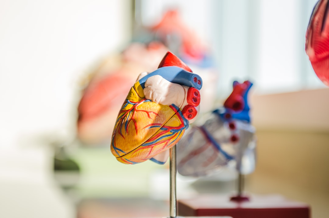
Heart valve developed at UC Irvine shines in early-stage preclinical testing
UC Irvine researchers designed and developed a minimally invasive replacement pulmonary heart valve. Created for pediatric patients, the device can be expanded as children grow, eliminating the need for multiple surgeries. The team successfully conducted laboratory and early-stage animal feasibility testing of the implant, crucial steps toward approval for human use. Irvine, Calif., June 23, 2025 — Researchers at the University of California, Irvine have successfully performed preclinical laboratory testing of a replacement heart valve intended for toddlers and young children with congenital cardiac defects, a key step toward obtaining approval for human use. The results of their study were published recently in the Journal of the American Heart Association. The management of patients with congenital heart disease who require surgical pulmonary valve replacement typically occurs between the ages of 2 and 10. To be eligible for a minimally invasive transcatheter pulmonary valve procedure, patients currently must weigh at least 45 pounds. For children to receive minimally invasive treatment, they must be large enough so that their veins can accommodate the size of a crimped replacement valve. The Iris Valve designed and developed by the UC Irvine team can be implanted in children weighing as little as 17 to 22 pounds and gradually expanded to an adult diameter as they grow. Research and development of the Iris Valve has been supported by the Eunice Kennedy Shriver National Institute of Child Health and Human Development; the National Heart, Lung, and Blood Institute; and the National Science Foundation. This funding has enabled benchtop fracture testing, which demonstrated the valve’s ability to be crimped down to a 3-millimeter diameter for transcatheter delivery and subsequently enlarged to 20 millimeters without damage, as well as six-month animal studies that confirmed successful device integration within the pulmonary valve annulus, showing valve integrity and a favorable tissue response. “We are pleased to see the Iris Valve performing as we expected in laboratory bench tests and as implants in Yucatan mini pigs, a crucial measure of the device’s feasibility,” said lead author Arash Kheradvar, UC Irvine professor of biomedical engineering. “This work represents the result of longstanding collaboration between our team at UC Irvine and Dr. Michael Recto at Children’s Hospital of Orange County built over several years of joint research and development.” Congenital heart defects affect about 1 percent of children born in the United States and Europe, with over 1 million cases in the U.S. alone. These conditions often necessitate surgical interventions early in life, with additional procedures required to address a leaky pulmonary valve and prevent right ventricular failure as children grow. The Iris Valve can be implanted via a minimally invasive catheter through the patient’s femoral vein. The Kheradvar group employed origami folding techniques to compress the device into a 12-French transcatheter system, reducing its diameter to no more than 3 millimeters. Over time, the valve can be balloon-expanded up to its full 20-millimeter diameter. This implantation method, along with the ability to begin treatment earlier in very young patients, helps mitigate the risk of complications from delayed care and reduces the need for multiple surgeries in this vulnerable population. “Once the Iris Valve comes to fruition, it will save hundreds of children at least one operation – if not two – throughout the course of their lives,” said Recto, an interventional pediatric cardiologist at CHOC who’s also a clinical professor of pediatrics at UC Irvine. “It will save them from having to undergo surgical pulmonary valve placement, as the Iris Valve is delivered via a small catheter in the vein and can be serially dilated to an adult diameter and also facilitate the future placement of larger transcatheter pulmonary valves – with sizes greater than 20 millimeters, like the Melody, Harmony and Sapien devices – if needed.” Kheradvar said that the next phase of preclinical testing of the Iris Valve is funded by the Brett Boyer Foundation, which is committed to supporting research into treatments for congenital heart disease. “We are actively engaged with the U.S. Food and Drug Administration to define and carry out the required experiments and documentation for first-in-human authorization of the Iris Valve,” Kheradvar said. “Our team is urgently advancing the Iris Valve through preclinical studies to enable its clearance for first-in-human use. This is a critical step toward providing toddlers – who currently have no viable minimally invasive treatment until they reach the 45-pound threshold – with a much-needed option.” First co-author Nnaoma Agwu, a biomedical engineering Ph.D. candidate at UC Irvine, said: “The development of the Iris Valve required a strong and knowledgeable team that understood the clinical and mechanical design requirements. This accomplishment would not have been possible without the collaboration of talented clinicians, veterinarians and engineers. With this milestone reached, we are rigorously advancing the Iris Valve’s development, setting our sights on human clinical trials.” Joining Kheradvar, Recto and Agwu as co-authors of the article in Journal of the American Heart Association were Daryl Chau, a recent UC Irvine master’s graduate; Gregory Kelley and Tanya Burney, both research specialists at UC Irvine, with Burney also affiliated with the Beckman Laser Institute; Ekaterina Perminov, a clinical veterinarian with UC Irvine’s University Laboratory Animal Resources; and Christopher Alcantara, a radiology technician at CHOC. About UC Irvine’s Brilliant Future campaign: Publicly launched on Oct. 4, 2019, the Brilliant Future campaign aims to raise awareness and support for the university. By engaging 75,000 alumni and garnering $2 billion in philanthropic investment, UC Irvine seeks to reach new heights of excellence in student success, health and wellness, research and more. The Samueli School of Engineering plays a vital role in the success of the campaign. Learn more by visiting https://brilliantfuture.UC Irvine.edu/the-henry-samueli-school-of-engineering About the University of California, Irvine: Founded in 1965, UC Irvine is a member of the prestigious Association of American Universities and is ranked among the nation’s top 10 public universities by U.S. News & World Report. The campus has produced five Nobel laureates and is known for its academic achievement, premier research, innovation and anteater mascot. Led by Chancellor Howard Gillman, UC Irvine has more than 36,000 students and offers 224 degree programs. It’s located in one of the world’s safest and most economically vibrant communities and is Orange County’s second-largest employer, contributing $7 billion annually to the local economy and $8 billion statewide. For more on UC Irvine, visit www.uci.edu. Media access: Radio programs/stations may, for a fee, use an on-campus studio with a Comrex IP audio codec to interview UC Irvine faculty and experts, subject to availability and university approval. For more UC Irvine news, visit news.uci.edu. Additional resources for journalists may be found at https://news.uci.edu/media-resources.

Taking discoveries to the real world for the benefit of human health
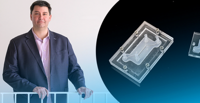
It takes about a decade and a lot of money to bring a new drug to market—between $1 billion to $2 billion, in fact. University of Delaware inventor Jason Gleghorn wants to change that. At UD, Gleghorn is developing leading-edge microfluidic tissue models. The devices are about the size of two postage stamps, and they offer a faster, less-expensive way to study disease and to develop pharmaceutical targets. These aren’t tools he wants to keep just for himself. No, Gleghorn wants to put the patented technology he’s developing in the hands of other experts, to advance clinical solutions in women’s health, maternal-fetal health and pre-term birth. His work also has the potential to improve understanding of drug transport in the female reproductive tract, placenta, lung and lymph nodes. Gleghorn, an associate professor of biomedical engineering, was named to the first cohort of Innovation Ambassadors at UD, as part of the University’s effort to foster and support an innovation culture on campus. Below, he shares some of what he’s learned about translating research to society. Q: What is the problem that you are trying to address? Gleghorn: A lot of disease has to do with disorganization in the body’s normal tissue structure. My lab makes microfluidic tissue models, called organ-on-a-chip models, that have super-tiny channels about the thickness of a human hair, where we can introduce very small amounts of liquid, including cells, to represent an organ in the human body. This can help us study and understand the mechanism of how things work in the body (the biology) or help us do things like drug screening to test therapeutic compounds for treating disease. And while these little microfluidic devices can do promising things, the infrastructure required to make the system work often restricts their use to high-end labs. We want to democratize the techniques and technology so that nonexperts can use it. To achieve this, we changed the way we make these devices, so that they are compatible with standard manufacturing, which means we can scale them and create them much easier. Gleghorn: One of the problems with drug screening, in general, is that animal model studies don’t always represent human biology. So, when we’re using animal models to test new drugs — which have been the best tool we have available — the results are not always apples to apples. Fundamentally, our microfluidic devices can model what happens in humans … we can plug in the relevant human components to understand how the mechanism is working and then ask questions about what drives those processes and identify targets for therapies to prevent the dysfunction. Q: What is innovative about this device? Gleghorn: The innovation part is this modularity — no one makes these devices this way. The science happens on the tiny tissue model insert, which is sandwiched between two pieces of clear acrylic. This allows us to watch what’s happening on the tissue model insert in real time. Meanwhile, the outer shell’s clamshell design provides flexibility: if we’re studying lung tissue and we want to study the female reproductive tract, all we do is unscrew the outer shell and insert the proper tissue model that mimics the female reproductive tract and we’re off. We’ve done a lot of the engineering to make it very simple to operate and use, and adaptable to common lab tools that everyone has, to eliminate the need for financial investment in things like specialized clean rooms, incubators and pumps, etc., so the technology can be useful in regular labs or easily deployable to far-flung locations or countries. With a laser cutter and $500 worth of equipment, you could conceivably mass manufacture these things for maternal medicine in Africa, for example. Democratizing the technology so it is compatible and useful for even an inexperienced user aligns with the mission of my lab, which focuses on scaling the science and the innovation faster, instead of only a few specialized labs being a bottleneck to uncovering new mechanisms of disease and the development of therapies. We patented this modularity, the way to build these tiny microfluidic devices and the simplicity of how it's used as a tool set, through UD’s Office of Economic Innovation and Partnerships (OEIP). Q: How have you translated this work so far? Gleghorn: To date, we've taken this microfluidic system to nine different research labs across seven countries and four continents — including the United States, the United Kingdom, Australia, France, Belgium and South Africa. These labs are using our technology to study problems in women’s health and collecting data with it. We’re developing boot camps where researchers can come for two or three days to the University of Delaware, where we teach them how to use this device and they take some back with them. From a basic science perspective, there is high enthusiasm for the power of what it can tell you and its ease of use. As engineers, we think it's pretty cool that many other people are using our innovations for new discoveries. Q: What support and guidance have you received from the UD innovation ecosystem? Gleghorn: To do any of this work, you need partners that have various expertise and backgrounds. UD’s Office of Economic Innovation and Partnerships has built a strong team of professionals with expertise in different areas, such as how do you license or take something to patent, how do you make connections with the business community? OEIP is home to Delaware’s Small Business Development Center, which can help you think about business visibility in terms of startups. Horn Entrepreneurship has built out impressive programs for teaching students and faculty to think entrepreneurially and build mentor networks, while programs like the Institute for Engineering Driven Health and the NSF Accelerating Research Translation at UD provide gap funding to be able to do product development and to take the work from basic prototype to something that is more marketable. More broadly in Delaware is the Small Business Administration, the Delaware Innovation Space and regional grant programs and small accelerators to help Delaware innovators. Q: How have students in your lab benefited from engaging in innovation? Gleghorn: Undergraduate students in my lab have made hundreds of these devices at scale. We basically built a little manufacturing facility, so we have ways to sterilize them, track batches, etc. We call it “the foundry.” In other work, graduate students are engineering different components or working on specific system designs for various studies. The students see collaborators use these devices to discover new science and new discoveries. That's very rewarding as an engineer. Additionally, my lab focuses on building solutions that are useful in the clinic and commercially viable. As a result, we've had two grad students spin out companies related to the work we've been doing in the lab. Q: How has research translation positively impacted your work? Gleghorn: I started down this road maybe five years ago, seriously trying to think about how to translate our research findings. Being an entrepreneur, translating technology — it's a very different way to think about your work. And so that framework has really permeated most of the research that I do now and changed the way I think about problems. It has opened new opportunities for collaboration and for alternate sources of funding with companies. This has value in terms of taking the research that you're doing fundamentally and creating a measurable impact in the community, but it also diversifies your funding streams to work on important problems. And different viewpoints help you look at the work you do in new ways, challenging you to define the value proposition, the impact of your work.


Swimming in the deep: MSU research reveals sea lamprey travel patterns in Great Lakes waterways
Why this matters: Invasive sea lampreys prey on most species of large Great Lakes fish such as lake trout, brown trout, lake sturgeon, lake whitefish, ciscoes, burbot, walleye and catfish. These species are crucial to Great Lakes ecosystems and to the region’s fishing industry. Understanding how sea lampreys migrate can inform management and conservation strategies, such as developing methods to catch the invasive fish that don’t involve dams, which reduce river connectivity, or lampricide, a pesticide that some communities and groups prefer not to use. The Great Lakes fishing industry is worth $7 billion and provides 75,000 jobs to the region. Reducing the amount of sea lamprey in waters is crucial for the industry’s well-being and the economic vitality of the Great Lakes. How do you catch an invasive fish that’s solitary, nocturnal and doesn't feed on bait? Researchers in the Michigan State University College of Agriculture and Natural Resources are one step closer to figuring it out. In a study published in the Journal of Experimental Biology and funded by the Great Lakes Fishery Commission, Kandace Griffin, a fisheries and wildlife doctoral student, and Michael Wagner, professor in the MSU Department of Fisheries and Wildlife, found that sea lampreys — a parasitic fish considered an invasive species in the Great Lakes region of the U.S. — follow a clear pattern of staying in the deepest parts of a river. These findings are important for informing sea lamprey management strategies, conservation of fish species native to the Great Lakes and protecting the region’s $7 billion fishing industry and the 75,000 jobs it provides. “We wanted to know how sea lampreys are making their movement decisions when migrating,” Griffin said. “Are they guided by certain environmental cues? Are they moving through areas that are safer? How can we potentially exploit those decisions or maybe manipulate them into going somewhere that they don’t want to go, like pushing them into a trap.” The primary methods used to control sea lamprey are dams that block them from entering waterways and lampricide, a species-specific pesticide that targets lamprey larvae. “Dams create a lot of challenges for conserving river ecosystems: They block all the other fish that are moving up and down in the system. Even though lampricide is proven to be safe and effective, there are communities that are uncomfortable with its use going into the future,” Wagner said. “Figuring out the right way to fish sea lamprey would decrease its population, lower reproduction rates and provide managers with the opportunity to match their control tactics to the community’s needs.” To track lamprey movements, Griffin and Wagner used a method called acoustic telemetry, which involved using sound emitted from a surgically implanted tag to track the movement of 56 sea lampreys in the White River near Whitehall, Michigan. Griffin likened acoustic telemetry to GPS. “There’s a tag that emits sound and has a unique transmission with a unique identification code, so I know exactly which fish is going where,” she said. “The receivers are listening for that sound and then calculating the time it reaches each receiver. We used this information to triangulate the position of the sea lamprey and analyzed it to find out how they’re using the river’s environmental traits to make decisions on where to swim.” Of the 56 lampreys studied, 26 of them (46%), consistently chose the deepest quarter of the river. “For nearly 20 years we have been discovering how sea lampreys migrate along coasts and through rivers. Now, thanks to Kandace’s work, we know where their movement paths come together near a riverbank — the perfect place to install a trap or other fishing device,” Wagner said. “That knowledge can be used to find similar sites across the Great Lakes basin.” Right now, a fishing device designed to catch bottom-swimming, solitary, nonfeeding, nocturnal sea lamprey doesn’t exist. However, Wagner notes there are places around the world — including Indigenous communities in the U.S. — where people have fished migratory lampreys of various species for hundreds of years and could help inform the creation of such a mechanism. “We have recently had a proposal funded to scour the Earth in search of knowledge, both scientific and traditional, about how to capture migrating lampreys and similar fishes,” Wagner said. “We want to talk with the communities of people who have histories fishing these animals and use this information, along with other data we’ve gathered, to conceive a device that could be used to fish sea lampreys.” Griffin views the new intel on lamprey migration patterns as a way to inform fishing practices to complement some of the existing control methods. “Hopefully, we can use this as a supplemental control method to the use of the barriers or dams,” she said. “We have societal pressure to remove barriers to enhance river connectivity, and some barriers are failing. Open water trapping is another way that we could try to still combat the invasive sea lamprey problem here but also promote river connectivity and other conservation goals for other species.” Wagner shares the same perspective. “When a community, or the Great Lakes Fishery Commission, or the governments of Canada and the U.S. come in and say, ‘We’d really rather be able to control this river with something other than lampricide,’ we want to be able to be able to provide 360-degree solutions that specify where to fish, when to fish and how to fish using fully prototyped and tested equipment,” he said. “We want our science to help solve real-world problems.”


Nibir Dhar, Ph.D., director of the Convergence Lab Initiative and professor in the Department of Electrical and Computer Engineering, was recently appointed to the Virginia Microelectronics Center endowed chair. This position gives Dhar the opportunity to shape future scientists and engineers, as well as pursue breakthrough research at the College of Engineering. “It’s more than an academic role,” said Dhar. “It’s about preparing students for complex problems they’ll solve in industry and defense.” Dhar teaches semiconductor and infrared device courses while researching next-generation materials for real-world applications. He also explores AI’s ability to improve human-machine interactions. With his accomplished background and experience at national defense labs, Dhar bridges classroom theory with practical engineering challenges his students will face in their careers. “It feels incredible to be recognized this way. Virginia Commonwealth University truly values faculty who pour themselves into student success and university growth. What really drives me is knowing I’m helping build the next generation of problem-solvers. That’s where the real satisfaction comes from.” said Dhar. This promotion encourages Dhar to make bigger strides for research development that will transform both teaching methods and how technology advances in military and commercial sectors.


The Asian Needle Ant (Brachyponera chinensis) Found in Southern Louisiana
In Louisiana, there are several ant species that are capable of stinging besides the red imported fire ant (Solenopsis invicta), such as the elongate twig ant (Pseudomyrmex gracilis), Comanche harvester ant (Pogonomyrmex comanche) and several species in the subfamily Ponerinae. The Asian needle ant (ANA) (Brachyponera chinensis) joins the list and has been confirmed in the state. Recent reports on the Asian needle ant by Mississippi State University extension entomologist, Santos J. Portugal and other urban entomologists in the region spurred the authors at Louisiana State University to investigate the presence of ANA in their state. By happenstance, two citizen scientists had reported sightings of the ant on iNaturalist at two Louisiana parks in August 2024 and June 2025. Therefore, on June 17, 2025, an LSU entomologist visited one of the parks to ground-truth the citing by collecting the ant, as he had prior experience with it. The Asian needle ant is a termite specialist, preferentially feeding on them, often living in close proximity with termite colonies and inside damp wood. To collect the ants, water-soaked wood was located in a forested area, broken open, revealing ANA, and they did not react aggressively to the disturbance. The ants immediately grabbed immature larva and retreated into crevices, not bothering the collectors at all. Upon retrieval, an LSU entomologist used a microscope at 40x to 60x magnification and the dichotomous key authored by MacGown (2003) to confirm that the collected specimens were ANA. It is important to verify the identity of invasive species submitted on citizen scientist projects as the images may not be of sufficient quality to get a positive identification. The ANA was discovered in the U.S. in 1934 while individuals were researching Argentine ants (Linepithema humile). Since the introduction of the ANA, it has spread to many states within the U.S., ranging from Wisconsin to Texas to the east coast. ANAs are medium sized (about 5 mm long) and slender. The species originated from Asia. Queens are slightly larger (6.5 mm) and look similar in appearance to workers. ANAs are black to dark brown in coloration, with light brown legs, mandibles and antennae. To distinguish the ant from other look-alikes, ANA has a large single petiole node that extends above the thorax or alitrunk, and a shiny mesopluron on the side of the thorax. ANA colonies are typically small in numbers, up to a few thousand individuals in large colonies. They are polygynous, meaning they have multiple queens. ANAs use a unique foraging behavior, where the worker carries another worker to a food resource, then drops off the worker to assist in food transport. ANAs do not form mounds, but instead nest in damp, high humidity areas, such as rotting logs, void spaces, under rocks and in leaf litter. They are typically found in forested areas. They also form multiple colonies within an area, which is called polydomy. ANAs swarm during the spring and early summer, although this time range may vary for Louisiana. People typically encounter the ants when they are working with wet wood or digging in moist soil. This is when someone may potentially be stung, although they are not aggressive. The sting is reported similar to that of a honeybee. Individuals who are allergic to stings may have a life-threatening anaphylactic response if stung by the ant, which requires medical attention. Wearing gloves is adequate protection from ANA stings while working with rotten wood or soil in infested areas. People who are sensitive to other insect stings should be aware of the potential for ANA stings and carry an approved rescue device for severe allergenic responses. In addition to feeding on termites, the Asian needle ant will feed upon beetles, craneflies, springtails and native ants found in their preferred habitats. Because of their ability to prey upon native ants, they can impact native species that deposit seeds in the soil, thus reducing floral diversity. Therefore, ANA is capable of reducing both native animal and plant diversity in infested areas. Article originally posted here.


When Luis Quiroga-Nuñez, Ph.D was appointed director of Florida Tech’s Ortega Observatory and its primary tenant – a non-functioning, 32-inch telescope – in 2023, he decided it was time to provide astronomy students and others a window to space. The observatory is already a base for research across a spectrum of cosmic exploration through disciplines such as astronomy and astrophysics, heliophysics, planetary science and astrobiology. However, current students have yet to see the stars up close, as the aging telescope, commissioned in 2008, has sat dormant for the last several years. With restoration, the telescope could be a powerful tool to train students to use professional telescopes and make observations – critical skills that will help prepare them for their future careers. It soon became apparent, however, that this was no simple task. The restoration would necessitate reverse engineering on a large scale to even understand how to fix and upgrade the telescope, much less actually repair it. It would also, as Quiroga-Nuñez wisely recognized, be its own powerful educational opportunity, providing unique hands-on learning opportunities for students in the College of Engineering and Science. “We are an institute of technology. We have perfectly capable people, like these young students, ready to join hands-on projects, get crazy and start to be creative.” Luis Quiroga-Nuñez With various issues to tackle and eager to support home-grown expertise, Quiroga-Nuñez and Lee Caraway, Ph.D, an instructor in the department of electrical engineering and computer science, recruited students with varied backgrounds, from astronomy to electrical engineering and computer science. Students could apply what they learned in class and grow their portfolios with a real-world project, the sort of experiential learning that is a hallmark of a Florida Tech education. Some improvements have been made, but the project remains an exciting puzzle for students and faculty alike. Here’s how they are doing it. An Interdisciplinary Project In January 2023, Quiroga-Nuñez partnered with Caraway to rebuild the telescope from the inside out. They say the conversation started over lunch, sketching ideas on a napkin. With various issues to tackle and eager to support home-grown expertise, Caraway and Quiroga-Nuñez recruited students with varied backgrounds, from astronomy to engineering to computer science. “This is about as real-world as you can get without leaving school. We have this giant piece of technology that is not working. Figure out why,” said recent graduate Adrianna Agustin ’24, who helped update the telescope’s communication system. “All of those problem-solving skills will directly translate to wherever we go in the future.” The project’s multidisciplinary nature also boosts collaboration between both sides of the college. “We keep integrating different parts of the university and involving students in a project that we were blinded by,” Quiroga-Nuñez says. “We sit between the scientists and the engineers.” And there’s no shortage of tasks. In addition to the refurbishment, Quiroga-Nuñez and Caraway are also completing routine telescope maintenance, with students taking on adjacent projects around the observatory. With the telescope repair, each student is given their own task, such as redesigning a small clip that supports the dome’s electric current, reviewing the conditions of the finder’s lens or understanding how analog devices control the telescope’s focus. This allocation allows each student to claim their own individual contribution to the greater telescope puzzle. Opening a Time Capsule The telescope’s biggest issues were mechanical and electrical, all exacerbated by age. Its motors were decades old and naturally failing, Caraway said. These motors controlled the telescope’s right ascension and declination – essentially, its ability to move. The chaotic interior also involved multiple individual systems with dozens of wires. And the circuits controlling the motors, which dated back to the 1980s, were also failing due to age. As Caraway noted, his students are sweeping off “dust older than them.” “The technology back then simply did not exist to control the motors, run the diagnostics and make it all happen,” Caraway explained. “They’re not designed to run 30 years.” Additionally, the computer program that controlled the motors was outdated and did not meet to the university’s security requirements. Given all this, the team needed to develop a new communication system for the telescope, starting with the computer software. They decided instead of purchasing an upgraded computer system, they could build and program their own in-house from scratch. Next, once the new computer was up and running, it needed motors to command. Marisa Guerra ’24 worked on a senior design project involving a robotic arm whose motor structure was the same as the telescope’s. She crafted a blueprint for the telescope’s new motors using what she learned for her capstone project. At the same time, Agustin worked on developing a cleaner communication system between the computer to the motors. Her senior design research focused on electric vehicles and their internal circuit systems, and she could replicate something similar within the telescope – but not without digging through the decaying electronics first. “We had to reverse engineer and actually redraw the circuits, which was good practice because a lot of the time, for senior design at least, you don’t really have to design a new circuit. You are just kind of puzzle-piecing it together,” Agustin said. “But with this circuit, all of them were bad.” Using Guerra’s and Agustin’s senior design research, the team reprogrammed the telescope’s circuits. What once took 20 wires to operate now only takes two. They also reduced the weight of the telescope’s motors from 40 pounds to just 2 pounds. Once the communication system was finished, the team was just waiting for mobility. And on a day in Spring 2024, thanks to the refurbished system, they were able to create movement within the telescope for the first time in years. “I didn’t even know if that device could move internally,” Quiroga-Nuñez says. The moment was celebrated, but the team knew this success triggered a new challenge. It was time to tackle high astrometric precision – a crucial element of properly tracking movement in space. “We are pointing to tiny points in the sky. If we do not track that properly, we are going to be lost in the universe,” Quiroga-Nuñez says. The Value of Time Perfecting precise movement is expected to take some time, but that’s not a bad thing, Quiroga-Nuñez says. He believes that a lengthy timeline will offer more value in the long run because it will give even more students a chance to get involved. Besides, its primary purpose will be to teach students how to use a telescope and allow them to make observations and prepare for their future careers. Ultimately, Quiroga-Nuñez predicts that the telescope could pick up its first image from space in about a year if everything stays on track. However, the team still has a lot of ground within the telescope to uncover, with an unpredictable number of potential troubleshooting challenges. For example, while rebuilding the motor, they discovered that the internal mirror that illuminates the telescope’s visuals was in poor condition – it needed cleaning and new aluminum to reflect enough light to see the telescope’s imagery, Agustin explains. So, the team had to remove the mirror and ship it to New York for refurbishment – a process that took several months. Once the mirror is reinstalled, they can return to their quest for better precision. The mirror is just one example of unpredictability in reverse-engineering. Ultimately, dedicating more time to understanding and solving the unforeseen challenges allows more students to participate in the telescope’s journey, Quiroga-Nuñez says. “This is like a big Lego for them,” he says. “They are learning the process, and the students, I think, will have found a very valuable life experience.” If you're interested in connecting with Luis Quiroga-Nuñez, director of Florida Tech’s Ortega Observatory - simply contact Adam Lowenstein, Director of Media Communications at Florida Institute of Technology at adam@fit.edu to arrange an interview today.


How authorship language helped catch a domestic terrorist – new podcast
In the latest episode of Writing Wrongs, hosts Professor Tim Grant and Dr Nicci MacLeod interview Dr Isobelle Clarke to unravel a case where forensic linguistics helped track down and convict a dangerous individual. Episode three, Imposters Tending to the Wild with Dr Isobelle Clarke, dives into the chilling case of Nikolaos Karvounakis, a self-proclaimed anarchist who planted a viable explosive device in Princes Street Gardens, Edinburgh, in 2018. Karvounakis, a Greek national, evaded capture for years, hiding behind online anonymity and extremist rhetoric. However, forensic linguists stepped in to analyse his anonymous blog posts, revealing patterns in his language that ultimately helped Police Scotland link him to the crime. The case not only demonstrates how linguistic evidence can be a powerful forensic tool but also raises crucial questions about the role of language analysis in modern terrorism investigations. On 11 January 2018, a suspicious cardboard box was discovered in a public seating area in Edinburgh’s Princes Street Gardens. After a controlled explosion, investigators determined the device could have caused serious harm had it detonated. With no immediate leads, the investigation stalled - until an anonymous blog post surfaced, claiming responsibility for the attack. The post, written in both English and Spanish, was linked to an eco-anarchist group called Individualists Tending to the Wild, a Mexican-based extremist organisation advocating violent action against technological progress. Crucially, the post included an image of the bomb’s interior, a detail only the perpetrator or law enforcement could have known. Police Scotland sought the expertise of Professor Tim Grant, who analysed the text, producing a linguistic profile that suggested the writer was neither a native English nor Spanish speaker - but rather someone influenced by another language entirely. Two years later, police identified Nikolaos Karvounakis as a suspect. Using comparative authorship analysis, Professor Tim Grant compared his online writings - including song lyrics from his rock band - to the manifesto. By dissecting word patterns, grammatical structures and stylistic quirks, he established that Karvounakis was the likely author. This evidence -alongside forensic meteorology, which linked photos of clouds in Karvounakis’ blog posts to the same weather conditions on the day of the crime - was used to secure a warrant and seize computers containing known writings by Karvounakis. To eliminate inevitable bias that would result from having worked the case for more than two years, Professor Grant invited Dr Isabelle Clarke onto the case as an independent forensic linguist. Using a version of the General Imposters Method, a technique similar to a police lineup but for language, Dr Clarke confirmed that the writing style in the blog post was the closest to Karvounakis’ known writings. Police Scotland put the evidence in the case, including the linguistic evidence, to Karvounakis, and secured a guilty plea. In February 2022, Nikolaos Karvounakis was sentenced to over eight years in prison under the UK’s Terrorism Act. Tim Grant, professor of forensic linguistics at Aston University, said: “The case highlights the growing importance of forensic linguistics in solving crimes, particularly in an age where digital anonymity combines with extremist ideologies. “It also highlights the how different types of language analysis can assist as a case moves through different stages of investigation.” Dr Nicci MacLeod, deputy director of the Aston Institute for Forensic Linguistics, said: “This episode offers listeners a behind-the-scenes look at the forensic methods that expose deception, identify threats and ultimately bring criminals to justice.” Dr Isobelle Clarke, a lecturer in security and protection science at Lancaster University and one of the first graduates from the campus-based MA Forensic Linguistics programme at Aston University, said: “It was great to be back at Aston University talking about the Karvounakis case for the Writing Wrongs podcast. “It’s an interesting case to highlight, as it shows how different types of language analysis can help with police investigations.” Writing Wrongs is available on Spotify, Apple Podcasts and all major streaming platforms. Listeners are encouraged to subscribe, share and engage with the hosts by submitting their forensic linguistics questions. Whether it’s about this case or broader forensic linguistic techniques, Professor Grant and Dr MacLeod welcome inquiries from listeners.


3D-printed lung model helps researchers study aerosol deposition in the lungs
Treating respiratory diseases is challenging. Inhalable medicines depend on delivering particles to the right lung areas, which is complicated by factors like the drug, delivery method and patient variability, or even exposure to smoke or asbestos particles. University of Delaware researchers have developed an adaptable 3D lung model to address this issue by replicating realistic breathing maneuvers and offering personalized evaluation of aerosol therapeutics. “If it's something environmental and toxic that we're worried about, knowing how far and how deep in the lung it goes is important,” said Catherine Fromen, University of Delaware Centennial Associate Professor for Excellence in Research and Education in the Department of Chemical and Biomolecular Engineering. “If it's designing a better pharmaceutical drug for asthma or a respiratory disease, knowing exactly where the inhaled aerosol lands and how deep the medicine can penetrate will predict how well that works.”that can replicate realistic breathing maneuvers and offer personalized evaluation of aerosol therapeutics under various breathing conditions. Fromen and two UD alumni have submitted a patent application on the 3D lung model invention through UD’s Office of Economic Innovation and Partnerships (OEIP), the unit responsible for managing intellectual property at UD. In a paper published in the journal Device, Fromen and her team demonstrate how their new 3D lung model can advance understanding of how inhalable medications behave in the upper airways and deeper areas of the lung. This can provide a broader picture on how to predict the effectiveness of inhalable medications in models and computer simulations for different people or age groups. The researchers detail in the paper how they built the 3D structure and what they’ve learned so far. Valuable research tool The purpose of the lung is gas exchange. In practice, the lung is often approximated as the size of a tennis court that is exchanging oxygen and carbon dioxide with the bloodstream in our bodies. This is a huge surface area, and that function is critical — if your lungs go down, you're in trouble. Fromen described this branching lung architecture like a tree that starts with a trunk and branches out into smaller and smaller limbs, ranging in size from a few centimeters in the trachea to about 100 microns (roughly the combined width of two hairs on your head) in the lung’s farthest regions. These branches create a complex network that filters aerosols as they travel through the lung. Just as tree branches end in leaves, the lung’s branches culminate in delicate, leaf-like structures called alveoli, where gases are exchanged. “Those alveoli in the deeper airways make the surface area that provides this necessary gas exchange, so you don't want environmental things getting in there where they can damage these sensitive, finer structures,” said Fromen, who has a joint appointment in biomedical engineering. Mimicking the complex structure and function of the lung in a lab setting is inherently challenging. The UD-developed 3D lung model is unique in several ways. First, the model breathes in the same cyclic motion as an actual lung. That’s key, Fromen said. The model also contains lattice structures to represent the entire volume and surface area of a lung. These lattices, made possible through 3D printing, are a critical innovation, enabling precise design to mimic the lung's filtering processes without needing to recreate its full biological complexity. “There's nothing currently out there that has both of these features,” she explained. “This means that we can look at the entire dosage of an inhaled medicine. We can look at exposure over time, and we can capture what happens when you inhale the medication and where the medicine deposits, as well as what gets exhaled as you breathe.” The testing process Testing how far an aerosol or environmental particle travels inside the 3D lung model is a multi-step process. The exposure of the model to the aerosol only takes about five minutes, but the analysis is time-consuming. The researchers add fluorescent molecules to the solution being tested to track where the particles deposit inside the model’s 150 different parts. “We wash each part and rinse away everything that deposits. The fluorescence is just a molecule in the solution. When it deposits, we know the concentration of that, so, when we rinse it out, we can measure how much fluorescence was recovered,” Fromen said. This data allows them to create a heat map of where the aerosols deposit throughout the lung model’s airways, which then can be validated against benchmarked clinical data for where such aerosols would be expected to go in a human under similar conditions. The team’s current model matches a healthy person under sitting/breathing conditions for a single aerosol size, but Fromen’s team is working to ensure the model is versatile across a much broader range of conditions. “An asthma attack, exercise, cystic fibrosis, chronic obstructive pulmonary disorder (COPD) — all those things are going to really affect where aerosols deposit. We want to make sure our model can capture those differences,” Fromen said. The ability to examine disease features like airway narrowing or mucus buildup could lead to more personalized care, such as tailored medication doses or redesigned inhalers. Currently, inhaled medicines follow a one-size-fits-all approach, but the UD-developed model offers a tool to address these issues and understand why many inhaled medicines fail clinical trials.

The new device is designed to reduce the risk of injuries when medicines being delivered into a vein enter the surrounding tissues It detects this problem at the earliest stages, before it is visible to the human eye The project is being supported by SPARK The Midlands at Aston University, a network to support technology development for unmet clinical needs. Clinicians at Birmingham Women’s and Children's NHS Foundation Trust (BWC) have joined with academics at Aston University to create an innovative sensor to reduce the risk of injuries caused when drugs being delivered into a vein enter the surrounding tissue. This complication, called extravasation, can cause harm and, in the most severe cases, life-changing injuries and permanent scarring. It happens most often when infusing medicines into peripheral intravenous (IV) devices, such as a cannula, but can also occur when infusing into a central venous access device. By joining together, BWC and Aston University are combining clinical, academic and engineering expertise to create a sensor that can detect extravasation at its earliest stages. Karl Emms, lead nurse for patient safety at BWC, said: “We've done lots of work across our Trust that has successfully reduced incidents. While we've made fantastic progress, there is only so much we can do as early signs of extravasation can be difficult to detect with the human eye. “The next step is to develop a technology that can do what people can't - detection as it happens. This will make a huge impact on outcomes as the faster we can detect extravasation, the less likely it is that it will cause serious harm.” The focused work to date addressing the issue has recently been recognised by the Nursing Times Awards 2024, winning the Patient Safety Improvement title for this year. This new project is supported by SPARK The Midlands, a network at Aston University dedicated to providing academic support to advance healthcare research discoveries in the region. SPARK The Midlands is the first UK branch of Stanford University's prestigious global SPARK programme. It comes as a result of Aston University’s active involvement in the delivery of the West Midlands Health Tech Innovation Accelerator (WMHTIA) – a government-funded project aimed at helping companies drive their innovations towards market success. The SPARK scheme helps to provide mentorship and forge networks between researchers, those with technical and specialist knowledge and potential sources of funding. SPARK members have access to workshops led by industry experts, covering topics such as medical device regulations, establishing good clinical trials, and creating an enticing target product profile to engage future funders. Luke Southan, head of research commercialisation at Aston University and SPARK UK director, said: “I was blown away when Karl first brought this idea to me. I knew we had to do everything we could to make this a reality. This project has the potential to transform the standard of care for a genuine clinical need, which is what SPARK is all about.” Work on another potentially transformative project has also begun as the team are working to develop a medical device that detects the position of a nasogastric feeding tube. There is a risk of serious harm and danger to life if nasogastric tubes move into the lungs, rather than the stomach, and feed is passed through them. Emms explained: “pH test strips can usually detect nasogastric tube misplacement, but some children undergoing treatment can have altered pH levels in the stomach. This means this test sometimes does not work. “A medical device that can detect misplacement can potentially stop harm and fatalities caused by these incidents.” SPARK will bring together engineers, academics and clinicians for both projects to develop the devices for clinical trial, with a goal of the technologies being ready for clinical use in three to five years. Southan said: “BWC is one of our first partners at SPARK and we're really excited to work with them to make a vital impact on paediatric healthcare in the Midlands and beyond." Notes to editors About Aston University For over a century, Aston University’s enduring purpose has been to make our world a better place through education, research and innovation, by enabling our students to succeed in work and life, and by supporting our communities to thrive economically, socially and culturally. Aston University’s history has been intertwined with the history of Birmingham, a remarkable city that once was the heartland of the Industrial Revolution and the manufacturing powerhouse of the world. Born out of the First Industrial Revolution, Aston University has a proud and distinct heritage dating back to our formation as the School of Metallurgy in 1875, the first UK College of Technology in 1951, gaining university status by Royal Charter in 1966, and becoming the Guardian University of the Year in 2020. Building on our outstanding past, we are now defining our place and role in the Fourth Industrial Revolution (and beyond) within a rapidly changing world. For media inquiries in relation to this release, contact Helen Tunnicliffe, Press and Communications Manager, on (+44) 7827 090240 or email: h.tunnicliffe@aston.ac.uk About Birmingham Women’s and Children’s NHS Foundation Trust Birmingham Women’s and Children’s NHS Foundation Trust (BWC) brings together the very best in paediatric and women’s care in the region and is proud to have many UK and world-leading surgeons, doctors, nurses, midwives and other allied healthcare professionals on its team. Birmingham Children’s Hospital is the UK’s leading specialist paediatric centre, caring for sick children and young people between 0 and 16 years of age. Based in the heart of Birmingham city centre, the hospital is a world leader in some of the most advanced treatments, complex surgical procedures and cutting-edge research and development. It is a nationally designated specialist centre for epilepsy surgery and also boasts a paediatric major trauma centre for the West Midlands, a national liver and small bowel transplant centre and a centre of excellence for complex heart conditions, the treatment of burns, cancer and liver and kidney disease. The hospital is also home to one of the largest Child and Adolescent Mental Health Services in the country, comprising of a dedicated inpatient Eating Disorder Unit and Acute Assessment Unit for regional referrals of children and young people with the most serious of problems (Tier 4) and Forward Thinking Birmingham community mental health service for 0- to 25-year-olds. Birmingham Women’s Hospital is a centre of excellence, providing a range of specialist health care services to over 50,000 women and their families every year from Birmingham, the West Midlands and beyond. As well as delivering more than 8,200 babies a year, it offers a full range of gynaecological, maternity and neonatal care, as well as a comprehensive genetics service, which serves men and women. Its Fertility Centre is one of the best in the country, while the fetal medicine centre receives regional and national referrals. The hospital is also an international centre for education, research and development with a research budget of over £3 million per year. It also hosts the national miscarriage research centre – the first of its kind in the UK - in partnership with Tommy’s baby charity. For interview requests please email the Communications Team on bwc.communications@nhs.net
